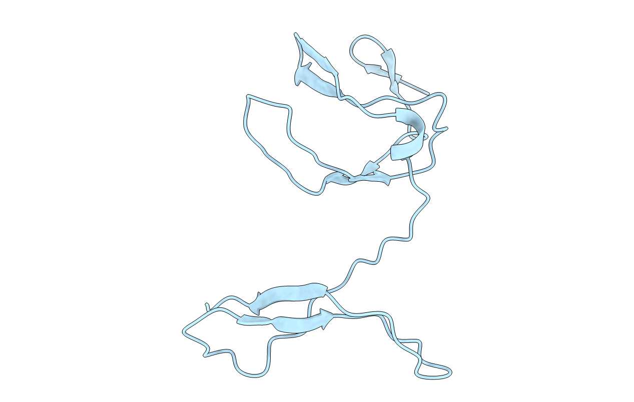
1zk9 image
Deposition Date 2005-05-02
Release Date 2005-05-10
Last Version Date 2023-08-23
Entry Detail
PDB ID:
1ZK9
Keywords:
Title:
NF-kB RelB forms an intertwined homodimer
Biological Source:
Source Organism:
Mus musculus (Taxon ID: 10090)
Host Organism:
Method Details:
Experimental Method:
Resolution:
2.18 Å
R-Value Free:
0.21
R-Value Work:
0.19
R-Value Observed:
0.19
Space Group:
P 31 2 1
Macromolecular Entities
Structures with similar UniProt ID
Protein Blast
Polymer Type:polypeptide(L)
Molecule:Transcription factor RelB
Gene (Uniprot):Relb
Chain IDs:A
Chain Length:110
Number of Molecules:1
Biological Source:Mus musculus
Primary Citation
NF-kappaB RelB Forms an Intertwined Homodimer.
Structure 13 1365 1373 (2005)
PMID: 16154093 DOI: 10.1016/j.str.2005.06.018

Abstact

The X-ray structure of the RelB dimerization domain (DD) reveals that the RelBDD assumes an unexpected intertwined fold topology atypical of other NF-kappaB dimers. All typical NF-kappaB dimers are formed by the association of two independently folded immunoglobulin (Ig) domains. In RelBDD, two polypeptides reconstruct both Ig domains in the dimer with an extra beta sheet connecting the two domains. Residues most critical to NF-kappaB dimer formation are invariant in RelB, and Y300 plays a positive role in RelBDD dimer formation. The presence of RelB-specific nonpolar residues at the surface removes several intradomain surface hydrogen bonds that may render the domain fold unstable. Intertwining may stabilize the RelBDD homodimer by forming the extra beta sheet. We show that, as in the crystal, RelB forms an intertwined homodimer in solution. We suggest that the transiently stable RelB homodimer might prevent its rapid degradation, allowing for heterodimer formation with p50 and p52.

Legend

Protein

Chemical

Disease

Primary Citation of related structures