1xhf image
Deposition Date 2004-09-18
Release Date 2005-05-17
Last Version Date 2024-11-06
Entry Detail
PDB ID:
1XHF
Keywords:
Title:
Crystal structure of the bef3-activated receiver domain of redox response regulator arca
Biological Source:
Source Organism:
Escherichia coli (Taxon ID: 562)
Host Organism:
Method Details:
Experimental Method:
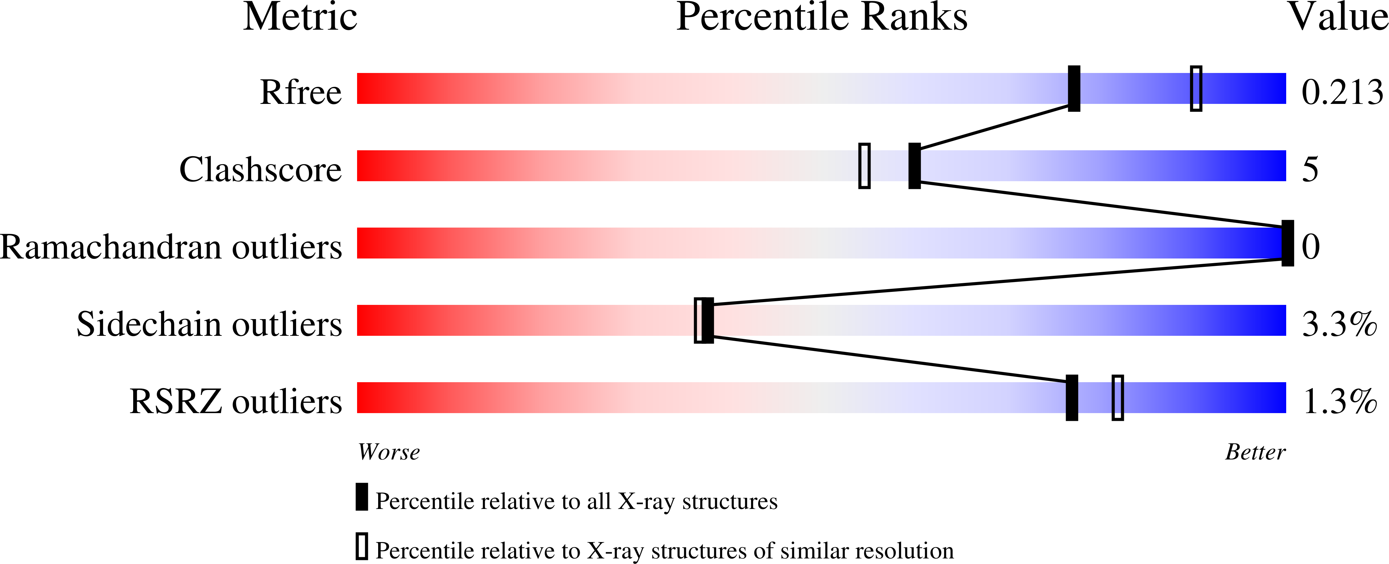
Resolution:
2.15 Å
R-Value Free:
0.21
R-Value Work:
0.18
R-Value Observed:
0.18
Space Group:
P 65 2 2
Macromolecular Entities
Structures with similar UniProt ID
Protein Blast
Polymer Type:polypeptide(L)
Molecule:Aerobic respiration control protein arcA
Gene (Uniprot):arcA
Mutagens:N123Q
Chain IDs:A, B
Chain Length:123
Number of Molecules:2
Biological Source:Escherichia coli
Modified Residue
Compound ID Chain ID Parent Comp ID Details 2D Image
MSE A MET SELENOMETHIONINE
Primary Citation
Structural Analysis and Solution Studies of the Activated Regulatory Domain of the Response Regulator ArcA: A Symmetric Dimer Mediated by the alpha4-beta5-alpha5 Face
J.Mol.Biol. 349 11 26 (2005)
PMID: 15876365 DOI: 10.1016/j.jmb.2005.03.059

Abstact

Escherichia coli react to changes from aerobic to anaerobic conditions of growth using the ArcA-ArcB two-component signal transduction system. This system, in conjunction with other proteins, regulates the respiratory metabolic pathways in the organism. ArcA is a member of the OmpR/PhoB subfamily of response regulator transcription factors that are known to regulate transcription by binding in tandem to target DNA direct repeats. It is still unclear in this subfamily how activation by phosphorylation of the regulatory domain of response regulators stimulates DNA binding by the effector domain and how dimerization and domain orientation, as well as intra- and intermolecular interactions, affect this process. In order to address these questions we have solved the crystal structures of the regulatory domain of ArcA in the presence and absence of the phosphoryl analog, BeF3-. In the crystal structures, the regulatory domain of ArcA forms a symmetric dimer mediated by the alpha4-beta5-alpha5 face of the protein and involving a number of residues that are highly conserved in the OmpR/PhoB subfamily. It is hypothesized that members of this subfamily use a common mechanism of regulation by dimerization. Additional biophysical studies were employed to probe the oligomerization state of ArcA, as well as its individual domains, in solution. The solution studies show the propensity of the individual domains to associate into oligomers larger than the dimer observed for the intact protein, and suggest that the C-terminal DNA-binding domain also plays a role in oligomerization.

Legend

Protein

Chemical

Disease

Primary Citation of related structures