1x3k image
Deposition Date 2005-05-09
Release Date 2005-05-24
Last Version Date 2024-10-23
Entry Detail
PDB ID:
1X3K
Title:
Crystal structure of a hemoglobin component (TA-V) from Tokunagayusurika akamusi
Biological Source:
Source Organism:
Method Details:
Experimental Method:
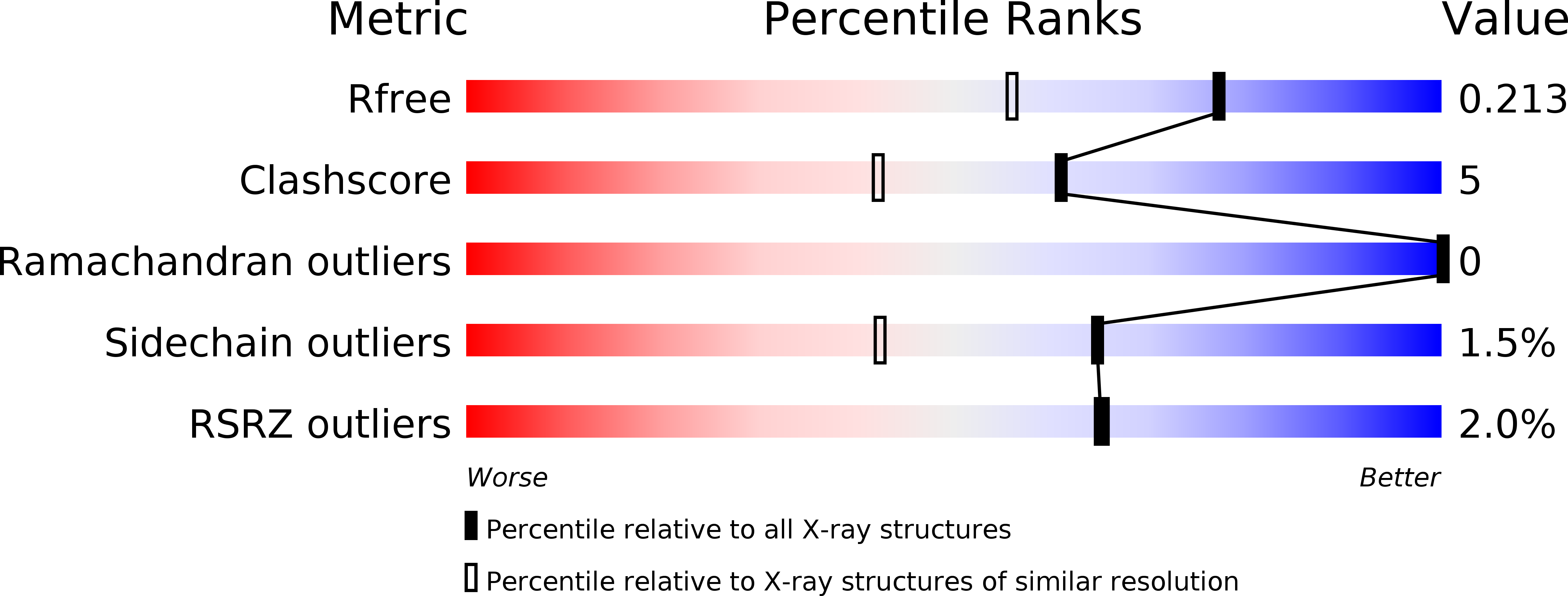
Resolution:
1.64 Å
R-Value Free:
0.21
R-Value Work:
0.19
Space Group:
P 21 21 2
Macromolecular Entities
Polymer Type:polypeptide(L)
Molecule:hemoglobin component V
Chain IDs:A
Chain Length:152
Number of Molecules:1
Biological Source:Tokunagayusurika akamusi
Ligand Molecules
Primary Citation
Crystal structures of two hemoglobin components from the midge larva Propsilocerus akamusi (Orthocladiinae, Diptera).
Gene 398 29 34 (2007)
PMID: 17590288 DOI: 10.1016/j.gene.2007.02.049

Abstact

The polymorphic components of hemoglobin (Hb) of the midge larva Propsilocerus akamusi were classified into two distinct types dependent on their spectroscopic properties, normal absorption (N) and low absorption (L). Analyses of the amino acid sequences of component VII (N-type Hb) and component V (L-type Hb) from P. akamusi indicated that one remarkable difference is the replacement of the distal histidine (His) with isoleucine (Ile) in component V. To clarify the structural differences between the two Hb components, we determined the crystal structures of components V and VII at resolutions of 1.64 A and 1.50 A, respectively. These crystal structures indicated a short additional helix comprising three amino acid residues at the C-terminal region in component V, and a typical globin fold including eight helices in component VII. Comparison of the heme regions of the Hb components suggests that the structural changes of the heme region in component V on ligation differ from that of usual Hb.

Legend

Protein

Chemical

Disease

Primary Citation of related structures