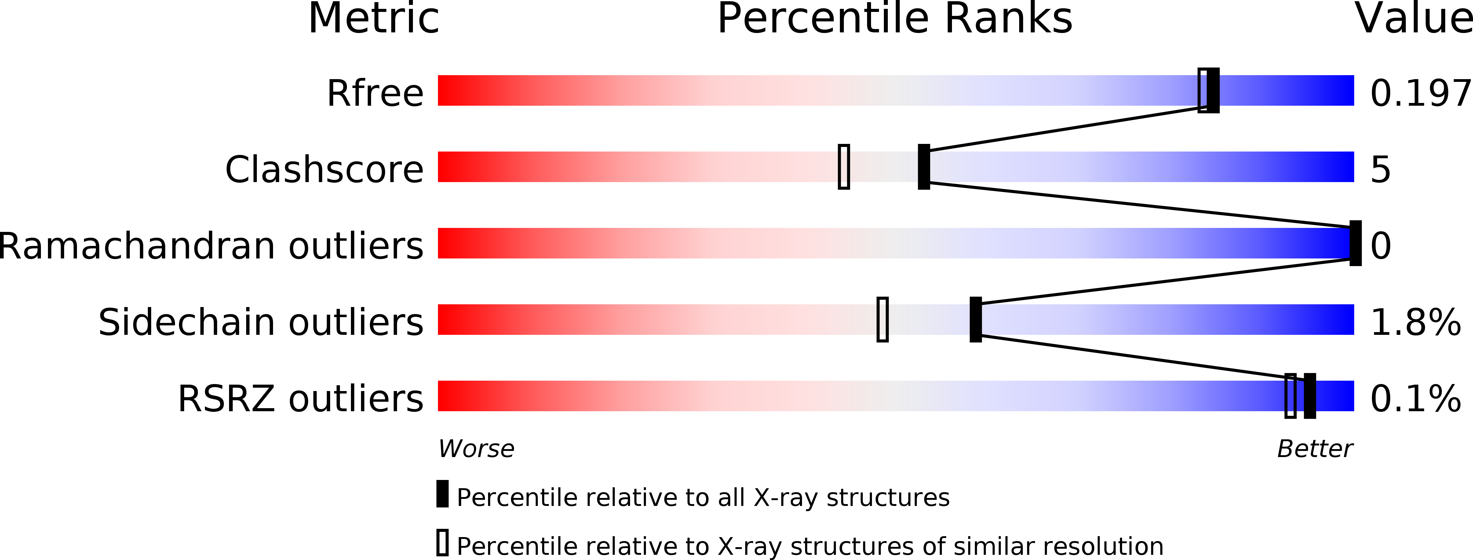
1va4 image
Deposition Date 2004-02-11
Release Date 2004-07-06
Last Version Date 2023-10-25
Entry Detail
PDB ID:
1VA4
Keywords:
Title:
Pseudomonas fluorescens aryl esterase
Biological Source:
Source Organism:
Host Organism:
Method Details:
Experimental Method:
Resolution:
1.80 Å
R-Value Free:
0.20
R-Value Work:
0.17
Space Group:
P 32
Macromolecular Entities
Polymer Type:polypeptide(L)
Molecule:Arylesterase
Gene (Uniprot):estF
Chain IDs:A, B, C, D, E, F
Chain Length:279
Number of Molecules:6
Biological Source:Pseudomonas fluorescens
Ligand Molecules
Primary Citation
Structure of an aryl esterase from Pseudomonas fluorescens.
Acta Crystallogr.,Sect.D 60 1237 1243 (2004)
PMID: 15213385 DOI: 10.1107/S0907444904010522

Abstact

The structure of PFE, an aryl esterase from Pseudomonas fluorescens, has been solved to a resolution of 1.8 A by X-ray diffraction and shows a characteristic alpha/beta-hydrolase fold. In addition to catalyzing the hydrolysis of esters in vitro, PFE also shows low bromoperoxidase activity. PFE shows highest structural similarity, including the active-site environment, to a family of non-heme bacterial haloperoxidases, with an r.m.s. deviation in 271 C(alpha) atoms between PFE and its five closest structural neighbors averaging 0.8 A. PFE has far less similarity (r.m.s. deviation in 218 C(alpha) atoms of 5.0 A) to P. fluorescens carboxyl esterase. PFE favors activated esters with small acyl groups, such as phenyl acetate. The X-ray structure of PFE reveals a significantly occluded active site. In addition, several residues, including Trp28 and Met95, limit the size of the acyl-binding pocket, explaining its preference for small acyl groups.

Legend

Protein

Chemical

Disease

Primary Citation of related structures