Deposition Date
2004-01-09
Release Date
2004-03-02
Last Version Date
2023-12-27
Entry Detail
PDB ID:
1V8K
Keywords:
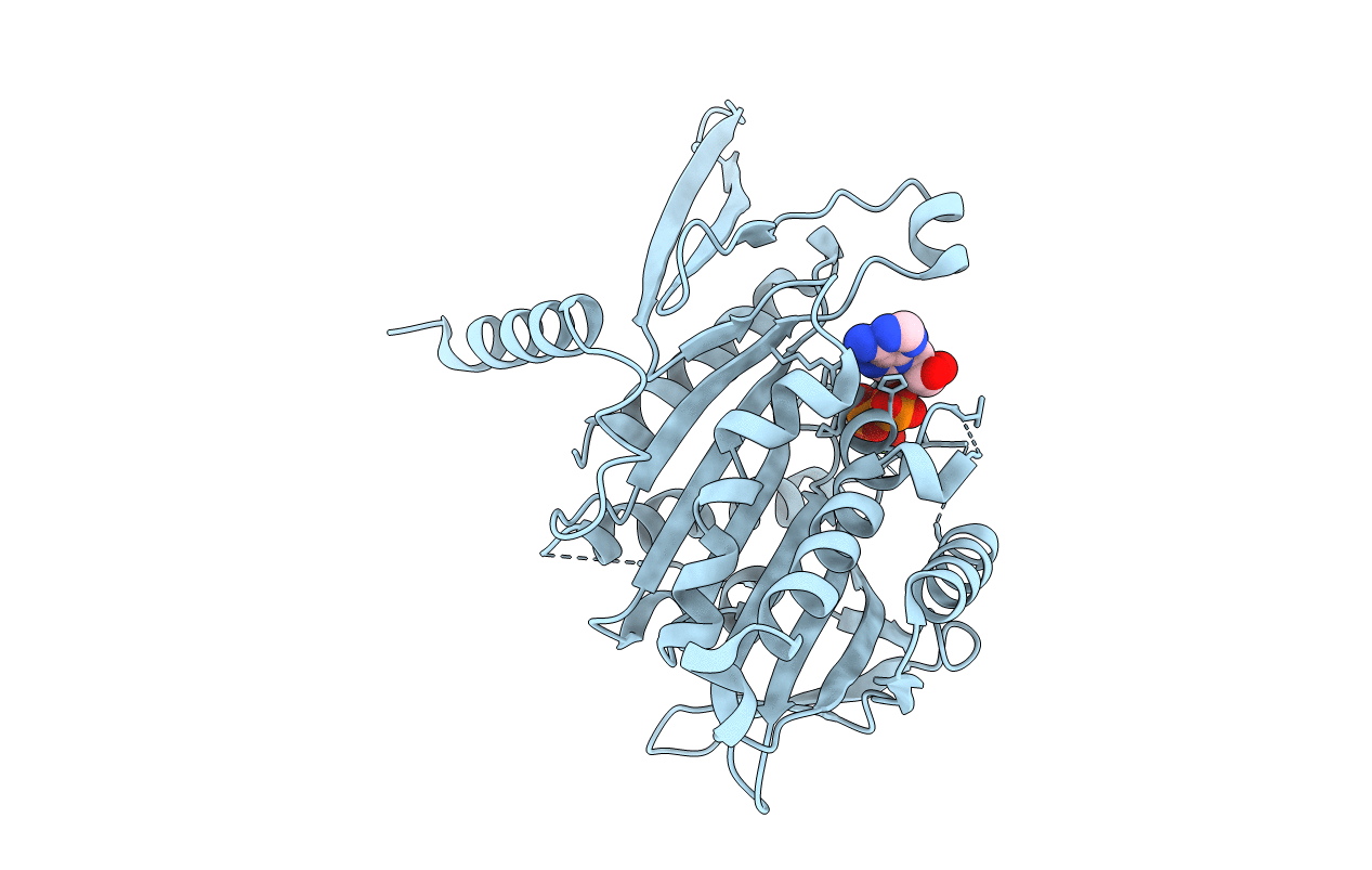
Title:
The Crystal Structure of the Minimal Functional Domain of the Microtubule Destabilizer KIF2C Complexed with Mg-AMPPNP
Biological Source:
Source Organism(s):
Mus musculus (Taxon ID: 10090)
Expression System(s):
Method Details:
Experimental Method:
Resolution:
2.25 Å
R-Value Free:
0.25
R-Value Work:
0.21
R-Value Observed:
0.21
Space Group:
C 2 2 2