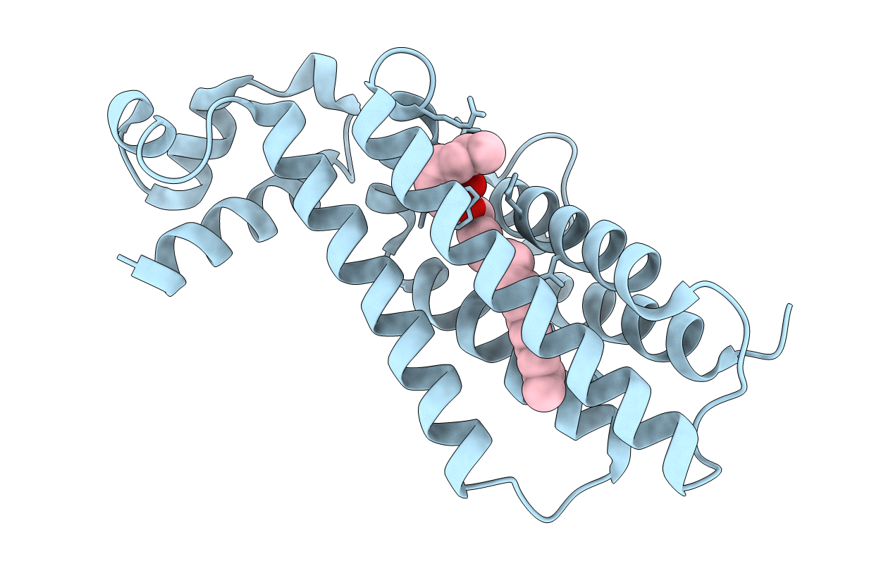
1u9n image
Deposition Date 2004-08-10
Release Date 2004-11-16
Last Version Date 2024-11-06
Entry Detail
PDB ID:
1U9N
Title:
Crystal structure of the transcriptional regulator EthR in a ligand bound conformation opens therapeutic perspectives against tuberculosis and leprosy
Biological Source:
Source Organism:
Host Organism:
Method Details:
Experimental Method:
Resolution:
2.30 Å
R-Value Free:
0.23
R-Value Work:
0.20
R-Value Observed:
0.20
Space Group:
P 41 21 2
Macromolecular Entities
Polymer Type:polypeptide(L)
Molecule:Transcriptional repressor EthR
Chain IDs:A
Chain Length:236
Number of Molecules:1
Biological Source:Mycobacterium tuberculosis
Modified Residue
Compound ID Chain ID Parent Comp ID Details 2D Image
MSE A MET SELENOMETHIONINE
Ligand Molecules
Primary Citation
Structure of EthR in a Ligand Bound Conformation Reveals Therapeutic Perspectives against Tuberculosis
Mol.Cell 16 301 307 (2004)
PMID: 15494316 DOI: 10.1016/j.molcel.2004.09.020

Abstact

Mycobacterium tuberculosis EthR is a repressor of ethA, a gene encoding a mono-oxygenase required for the activation of the prodrug ethionamide. Here we describe the X-ray crystal structure of EthR, a homodimer with an entirely helical structure showing similarities to TetR family members. Each monomer contained a fortuitous ligand identified as hexadecyl octanoate. The crystal structure of EthR purified in M. smegmatis revealed the presence of a comparable ligand. The binding of hexadecyl octanoate to EthR induces a conformational state incompatible with repressor function, which should lead to ethA derepression and consequently to an increased sensitivity to ethionamide and other thioamides. A related, more hydrophilic ketone was found to exhibit synergistic antimycobacterial effects when tested together with ethionamide, indicating that this strategy may help reduce the dosage of potent antibacterial compounds that otherwise are too toxic to be used as first-line drugs.

Legend

Protein

Chemical

Disease

Primary Citation of related structures