1slw image
Deposition Date 1996-02-07
Release Date 1996-07-11
Last Version Date 2024-10-30
Entry Detail
PDB ID:
1SLW
Title:
RAT ANIONIC N143H, E151H TRYPSIN COMPLEXED TO A86H ECOTIN; NICKEL-BOUND
Biological Source:
Source Organism:
Host Organism:
Method Details:
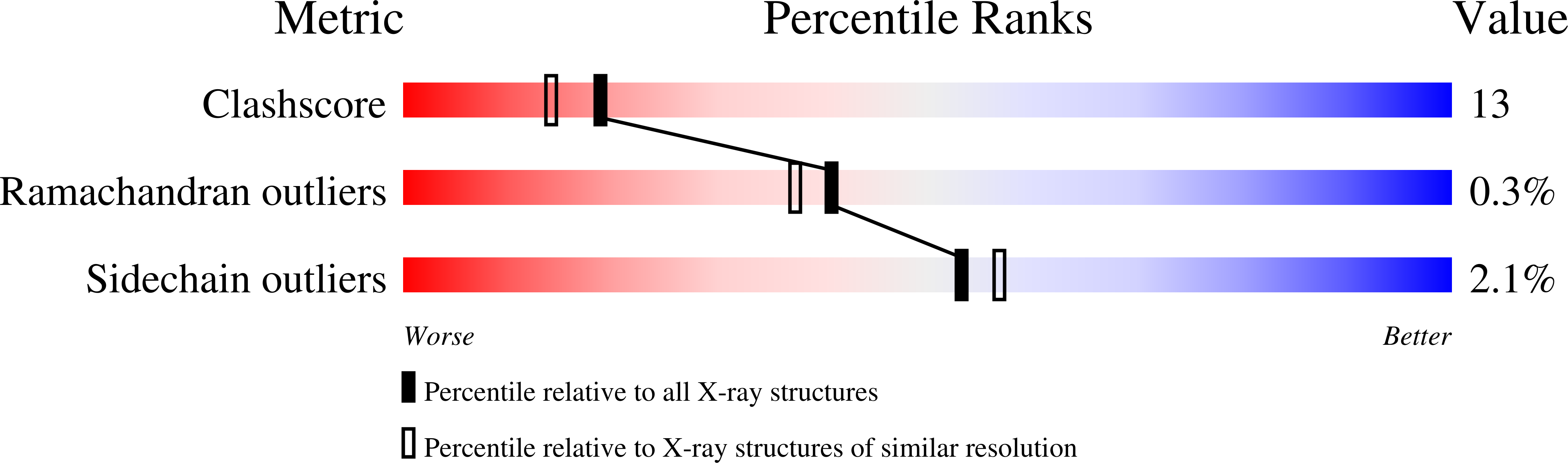
Experimental Method:
Resolution:
2.00 Å
R-Value Free:
0.25
R-Value Work:
0.17
R-Value Observed:
0.17
Space Group:
C 1 2 1
Macromolecular Entities
Polymer Type:polypeptide(L)
Molecule:ECOTIN
Gene (Uniprot):eco
Mutations:CHAIN A, A86H
Chain IDs:A
Chain Length:142
Number of Molecules:1
Biological Source:Escherichia coli
Polymer Type:polypeptide(L)
Molecule:ANIONIC TRYPSIN
Gene (Uniprot):Prss2
Mutations:CHAIN B, V121I, N143H, E151H
Chain IDs:B
Chain Length:223
Number of Molecules:1
Biological Source:Rattus norvegicus
Primary Citation
X-ray structures of a designed binding site in trypsin show metal-dependent geometry.
Biochemistry 35 5999 6009 (1996)
PMID: 8634241 DOI: 10.1021/bi9530200

Abstact

The three-dimensional structures of complexes of trypsin N143H, E151H bound to ecotin A86H are determined at 2.0 A resolution via X-ray crystallography in the absence and presence of the transition metals Zn2+, Ni2+, and Cu2+. The binding site for these transition metals was constructed by substitution of key amino acids with histidine at the trypsin-ecotin interface in the S2'/P2' pocket. Three histidine side chains, two on trypsin at positions 143 and 151 and one on ecotin at position 86, anchor the metals and provide extended catalytic recognition for substrates with His in the P2' pocket. Comparisons of the three-dimensional structures show the different geometries that result upon the binding of metal in the engineered tridentate site and suggest a structural basis for the kinetics of the metal-regulated catalysis. Of the three metals, the binding of zinc results in the most favorable binding geometry, not dissimilar to those observed in naturally occurring zinc binding proteins.

Legend

Protein

Chemical

Disease

Primary Citation of related structures