Deposition Date
2004-03-04
Release Date
2004-09-14
Last Version Date
2024-05-22
Entry Detail
PDB ID:
1SJQ
Keywords:
Title:
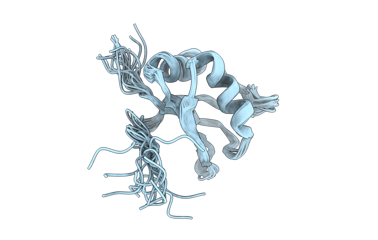
NMR Structure of RRM1 from Human Polypyrimidine Tract Binding Protein Isoform 1 (PTB1)
Biological Source:
Source Organism:
Homo sapiens (Taxon ID: 9606)
Host Organism:
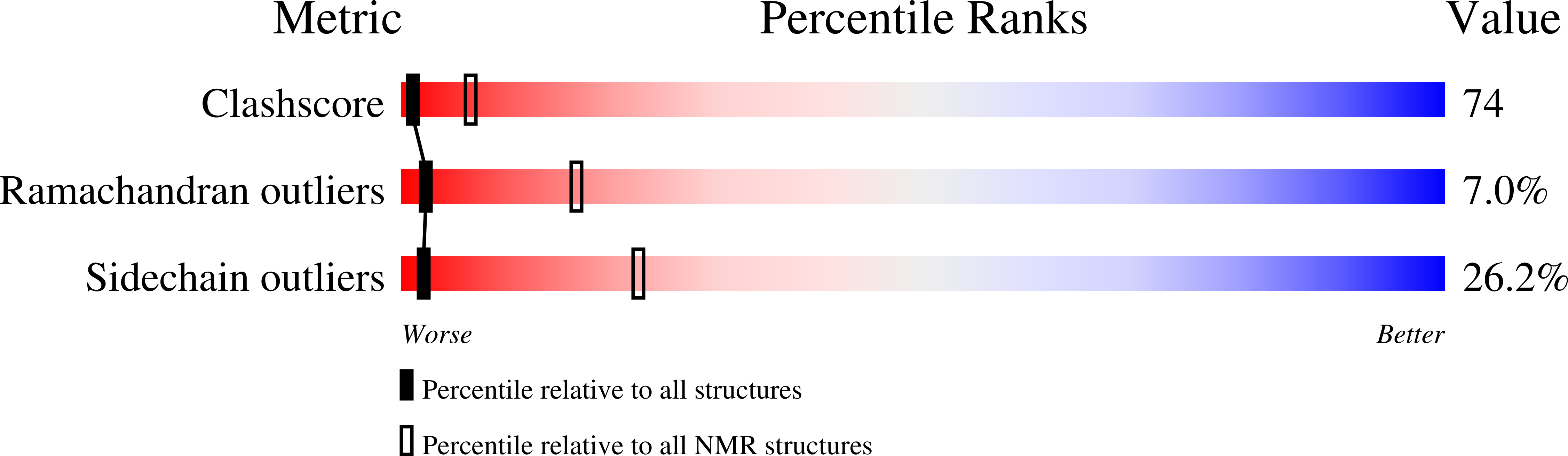
Method Details:
Experimental Method:
Conformers Calculated:
50
Conformers Submitted:
16
Selection Criteria:
structures with lowest energy