1sj1 image
Entry Detail
PDB ID:
1SJ1
Title:
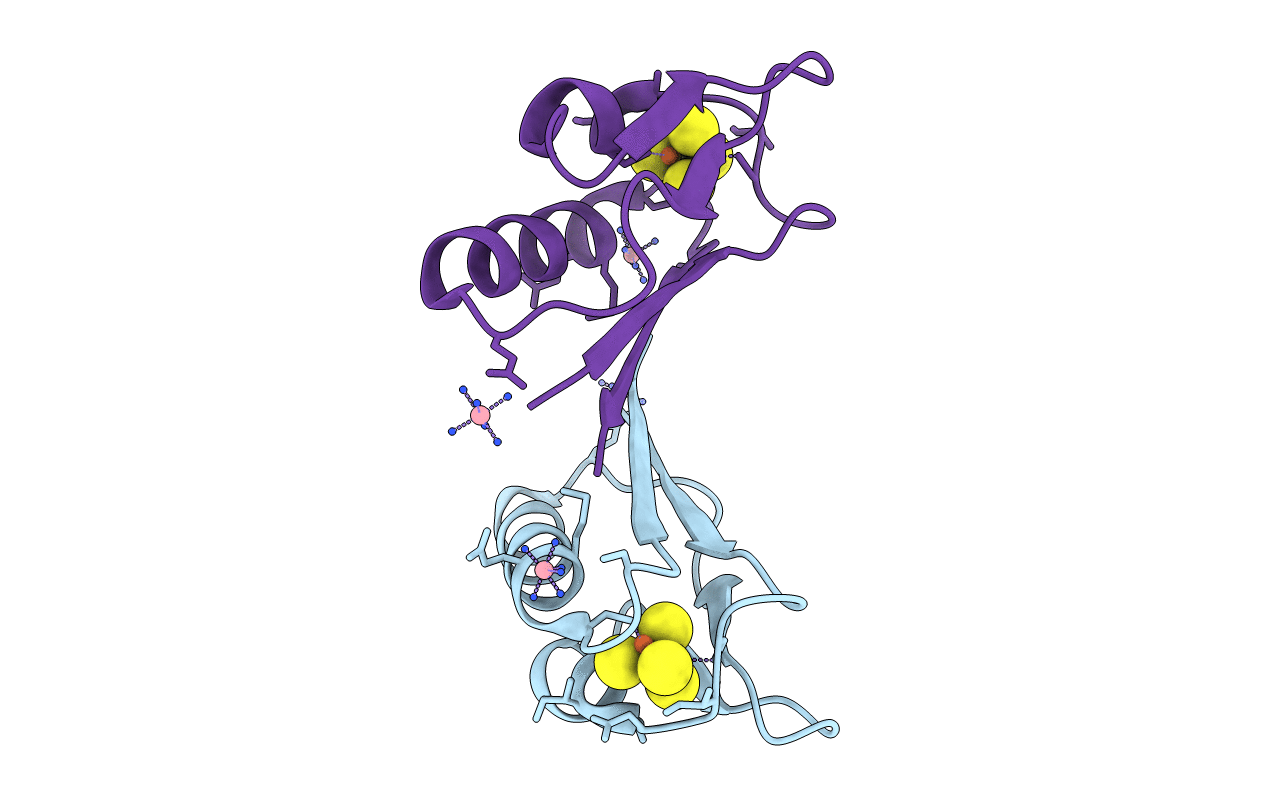
The 1.5 A Resolution Crystal Structure of [Fe3S4]-Ferredoxin from the hyperthermophilic Archaeon Pyrococcus furiosus
Biological Source:
Source Organism:
Host Organism:
PDB Version:
Deposition Date:
2004-03-02
Release Date:
2004-05-25
Method Details:
Experimental Method:
Resolution:
1.50 Å
R-Value Free:
0.22
R-Value Work:
0.19
R-Value Observed:
0.19
Space Group:
P 1 21 1
Macromolecular Entities
Polymer Type:polypeptide(L)
Description:Ferredoxin
Chain IDs:A, B
Chain Length:66
Number of Molecules:2
Biological Source:Pyrococcus furiosus
Primary Citation
The 1.5 A Resolution Crystal Structure of [Fe3S4]-Ferredoxin from the Hyperthermophilic Archaeon Pyrococcus furiosus
Biochemistry 43 5188 5194 (2004)
PMID: 15122884 DOI: 10.1021/bi049942x

Abstact

The structure of [Fe(3)S(4)]-ferredoxin from the hyperthermophilic archaeon Pyrococcus furiosus has been determined to 1.5 A resolution from a crystal belonging to space group P2(1) with two molecules in the asymmetric unit. The structure has been solved with molecular replacement by use of the ferredoxin from Thermotoga maritima. The fold is similar to that of related monocluster ferredoxins and contains two double-stranded antiparallel beta-sheets and two alpha-helices. The hydrophobic interaction between Trp2 and Tyr46 is confirmed, linking the C-terminus to the longer alpha-helix. The structure contains a double-conformation disulfide bond existing in a left-handed and a right-handed spiral conformation. The crystal packing reveals a beta-sheet interaction, which supports the suggestion that P. furiosus ferredoxin is a functional dimer. The extraordinary thermostability of P. furiosus ferredoxin is further discussed.

Legend

Protein

Chemical

Disease

Primary Citation of related structures