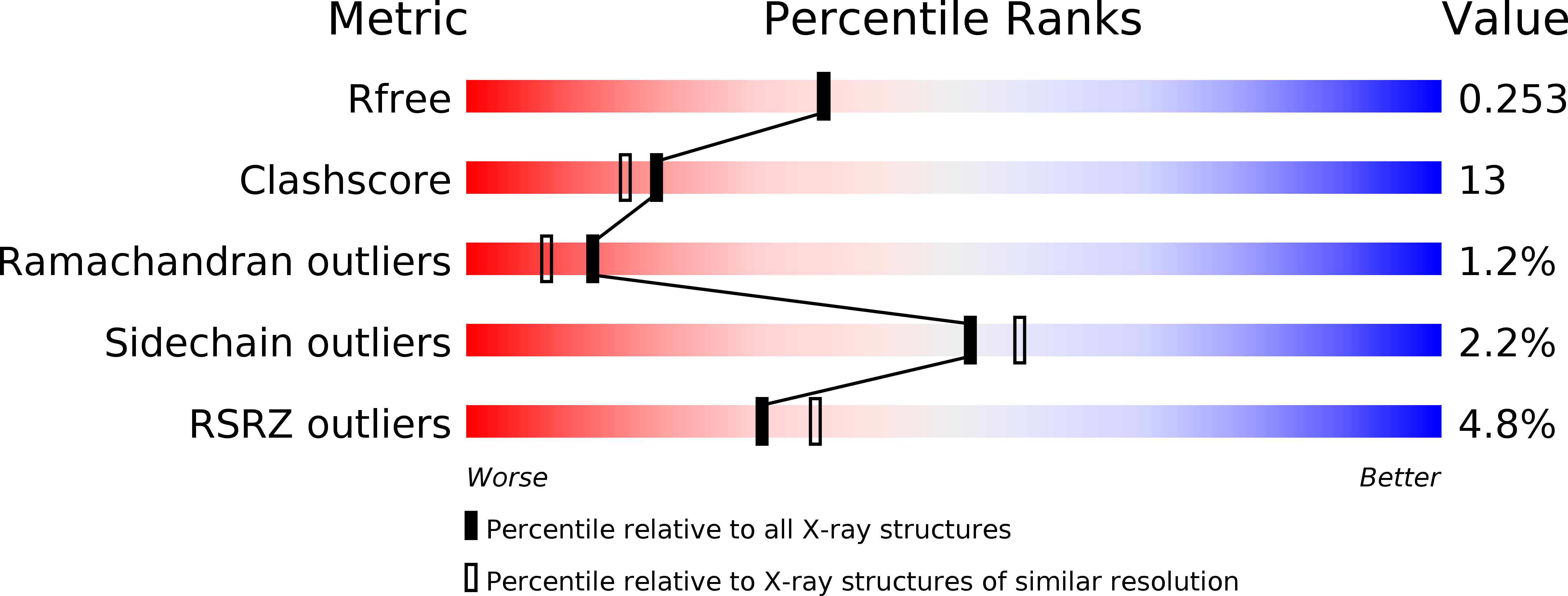
1s7m image
Deposition Date 2004-01-29
Release Date 2004-04-06
Last Version Date 2024-04-03
Entry Detail
PDB ID:
1S7M
Keywords:
Title:
Crystal Structure of HiaBD1
Biological Source:
Source Organism:
Method Details:
Experimental Method:
Resolution:
2.10 Å
R-Value Free:
0.25
R-Value Work:
0.21
R-Value Observed:
0.21
Space Group:
P 1 21 1
Macromolecular Entities
Structures with similar UniProt ID
Protein Blast
Polymer Type:polypeptide(L)
Molecule:Hia
Gene (Uniprot):hia
Chain IDs:A, B, C, D, E, F
Chain Length:174
Number of Molecules:6
Biological Source:Haemophilus influenzae
Primary Citation
Structural basis for host recognition by the Haemophilus influenzae Hia autotransporter.
Embo J. 23 1245 1256 (2004)
PMID: 15029242 DOI: 10.1038/sj.emboj.7600142

Abstact

Haemophilus influenzae is an important human pathogen that initiates infection by colonizing the upper respiratory tract. The H. influenzae Hia autotransporter is an adhesive protein that promotes adherence to respiratory epithelial cells. Hia adhesive activity resides in two homologous binding domains, called HiaBD1 and HiaBD2. These domains interact with the same host cell receptor, but bind with different affinities. In this report, we describe the crystal structure of the high-affinity HiaBD1 binding domain, which has a novel trimeric architecture with three-fold symmetry and a mushroom shape. The subunit constituents of the trimer are extensively intertwined. The receptor-binding pocket is formed by an acidic patch that is present on all three faces of the trimer, providing potential for a multivalent interaction with the host cell surface, analogous to observations with the trimeric tumor necrosis factor superfamily of proteins. Hia is a novel example of a bacterial trimeric adhesin and may be the prototype member of a large family of bacterial virulence proteins with a similar architecture.

Legend

Protein

Chemical

Disease

Primary Citation of related structures