1s4g image
Deposition Date 2004-01-16
Release Date 2004-06-08
Last Version Date 2024-11-13
Entry Detail
PDB ID:
1S4G
Keywords:
Title:
Somatomedin-B Domain of human plasma vitronectin.
Biological Source:
Source Organism:
Homo sapiens (Taxon ID: 9606)
Method Details:
Experimental Method:
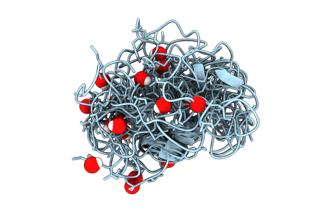
Conformers Calculated:
60
Conformers Submitted:
20
Selection Criteria:
structures with favorable non-bond energy
Macromolecular Entities
Polymer Type:polypeptide(L)
Molecule:Vitronectin
Gene (Uniprot):VTN
Chain IDs:A
Chain Length:51
Number of Molecules:1
Biological Source:Homo sapiens
[{'entity_id': ['1'], 'name': ['NA'], 'comp_id': ['NA'], 'asym_id': ['NA'], 'ndb_seq_num': ['1'], 'pdb_seq_num': ['1'], 'auth_seq_num': ['1'], 'pdb_mon_id': ['NA'], 'auth_mon_id': ['NA'], 'pdb_strand_id': ['NA'], 'pdb_ins_code': ['.']}]
Primary Citation
The solution structure of the N-terminal domain of human vitronectin: proximal sites that regulate fibrinolysis and cell migration
J.Biol.Chem. 279 29359 29366 (2004)
PMID: 15123712 DOI: 10.1074/jbc.M401279200

Abstact

The three-dimensional structure of an N-terminal fragment comprising the first 51 amino acids from human plasma vitronectin, the somatomedin B (SMB) domain, has been determined by two-dimensional NMR approaches. An average structure was calculated, representing the overall fold from a set of 20 minimized structures. The core residues (18-41) overlay with a root mean square deviation of 2.29 +/- 0.62 A. The N- and C-terminal segments exhibit higher root mean square deviations, reflecting more flexibility in solution and/or fewer long-range NOEs for these regions. Residues 26-30 form a unique single-turn alpha-helix, the locus where plasminogen activator inhibitor type-1 (PAI-1) is bound. This structure of this helix is highly homologous with that of a recombinant SMB domain solved in a co-crystal with PAI-1 (Zhou, A., Huntington, J. A., Pannu, N. S., Carrell, R. W., and Read, R. J. (2003) Nat. Struct. Biol. 10, 541-544), although the remainder of the structure differs. Significantly, the pattern of disulfide cross-links observed in this material isolated from human plasma is altogether different from the disulfides proposed for recombinant forms. The NMR structure reveals the relative orientation of binding sites for cell surface receptors, including an integrin-binding site at residues 45-47, which was disordered and did not diffract in the co-crystal, and a site for the urokinase receptor, which overlaps with the PAI-1-binding site.

Legend

Protein

Chemical

Disease

Primary Citation of related structures