Deposition Date
2003-12-17
Release Date
2004-05-11
Last Version Date
2023-08-23
Entry Detail
PDB ID:
1RWY
Keywords:
Title:
CRYSTAL STRUCTURE OF RAT ALPHA-PARVALBUMIN AT 1.05 RESOLUTION
Biological Source:
Source Organism(s):
Rattus norvegicus (Taxon ID: 10116)
Expression System(s):
Method Details:
Experimental Method:
Resolution:
1.05 Å
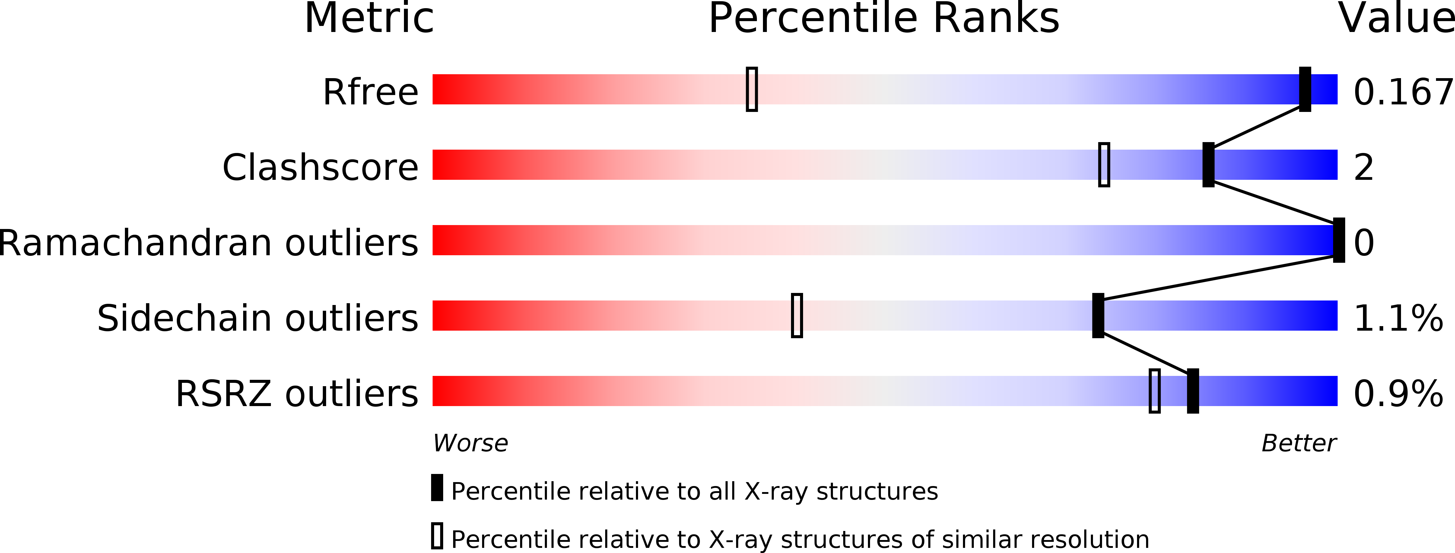
R-Value Free:
0.16
R-Value Observed:
0.13
Space Group:
P 21 21 21