Deposition Date
1999-07-01
Release Date
1999-10-08
Last Version Date
2024-10-30
Entry Detail
PDB ID:
1QUB
Keywords:
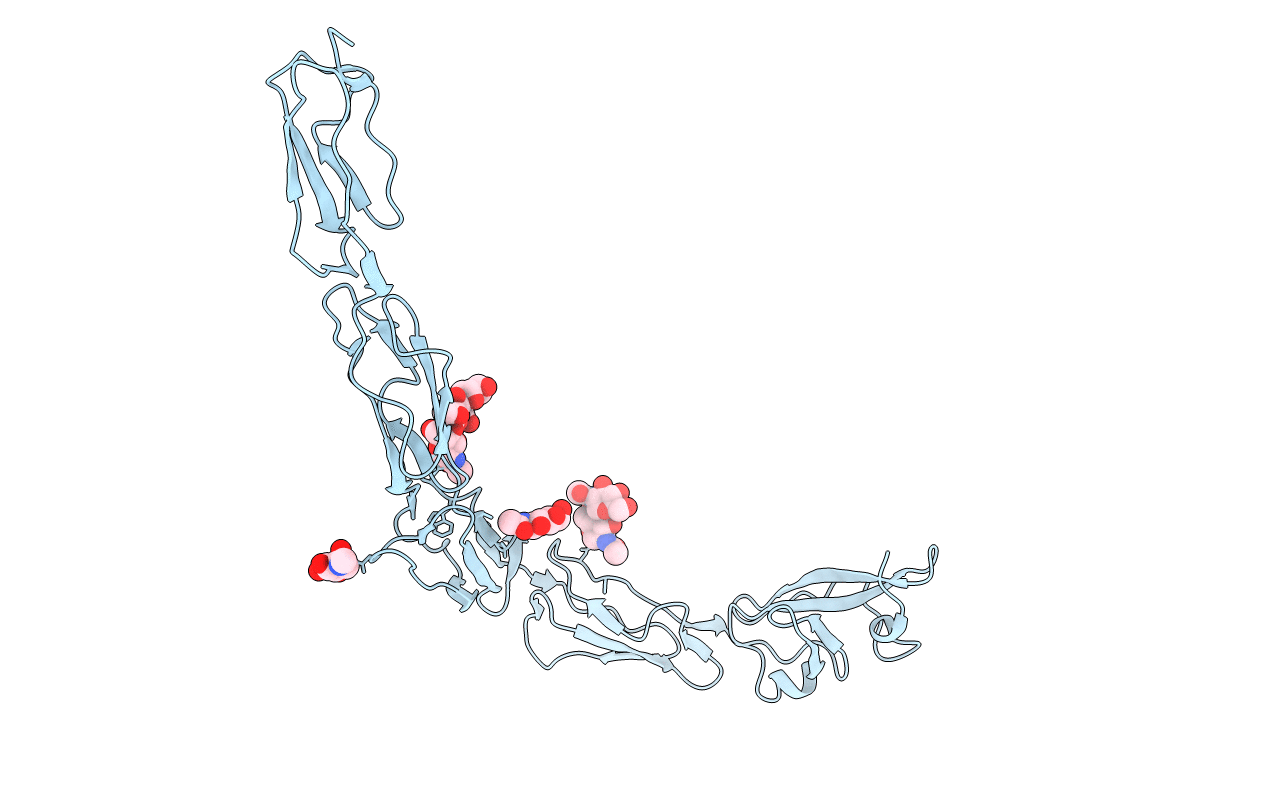
Title:
CRYSTAL STRUCTURE OF THE GLYCOSYLATED FIVE-DOMAIN HUMAN BETA2-GLYCOPROTEIN I PURIFIED FROM BLOOD PLASMA
Biological Source:
Source Organism(s):
Homo sapiens (Taxon ID: 9606)
Method Details:
Experimental Method:
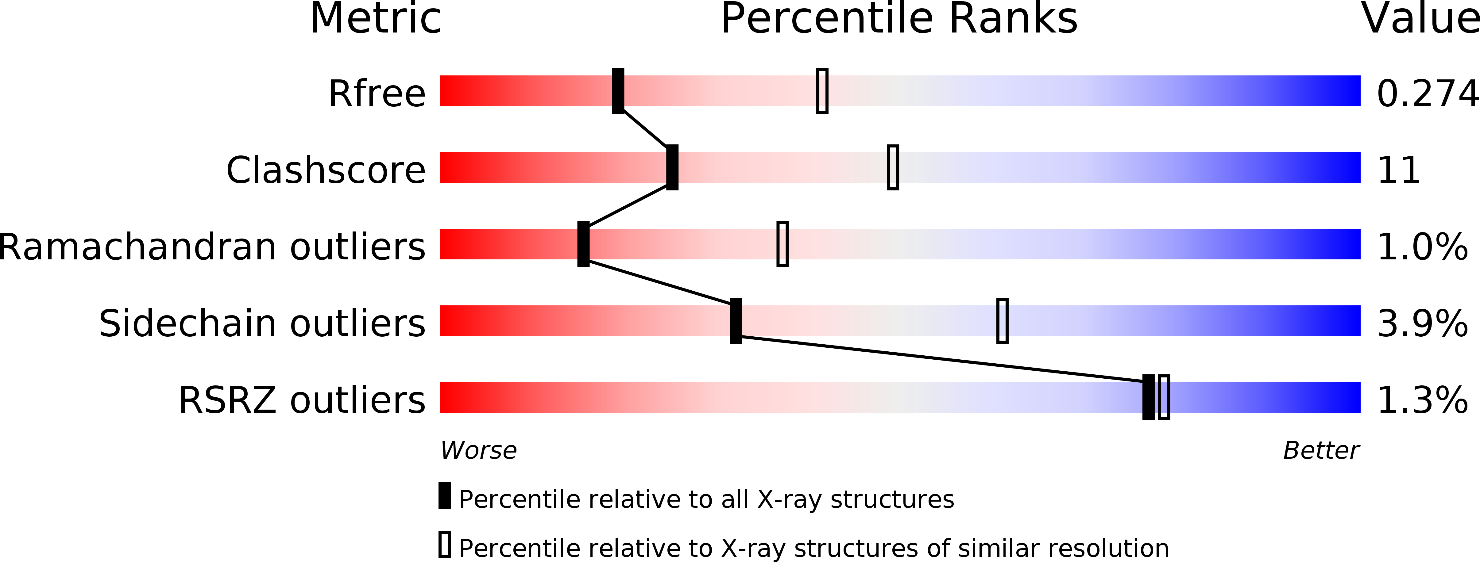
Resolution:
2.70 Å
R-Value Free:
0.26
R-Value Work:
0.24
R-Value Observed:
0.25
Space Group:
C 2 2 21