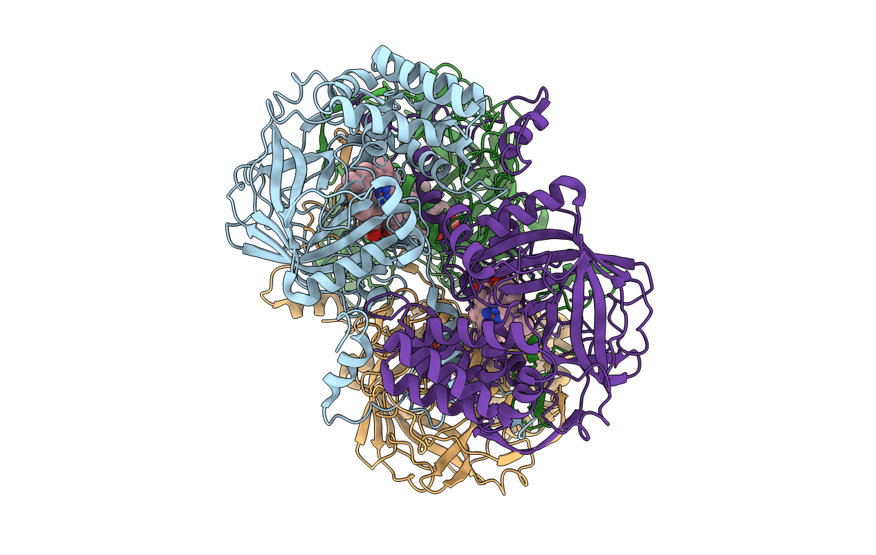
1qqw image
Deposition Date 1999-06-09
Release Date 1999-06-14
Last Version Date 2024-02-14
Entry Detail
PDB ID:
1QQW
Keywords:
Title:
CRYSTAL STRUCTURE OF HUMAN ERYTHROCYTE CATALASE
Biological Source:
Source Organism:
Homo sapiens (Taxon ID: 9606)
Method Details:
Experimental Method:
Resolution:
2.75 Å
R-Value Free:
0.27
R-Value Work:
0.20
R-Value Observed:
0.21
Space Group:
P 21 21 21
Macromolecular Entities
Structures with similar UniProt ID
Protein Blast
Polymer Type:polypeptide(L)
Molecule:CATALASE
Gene (Uniprot):CAT
Chain IDs:A, B, C, D
Chain Length:527
Number of Molecules:4
Biological Source:Homo sapiens
Ligand Molecules
Primary Citation
Structure of human erythrocyte catalase.
Acta Crystallogr.,Sect.D 56 241 245 (2000)
PMID: 10666617 DOI: 10.1107/S0907444999015930

Abstact

Catalase (E.C. 1.11.1.6) was purified from human erythrocytes and crystallized in three different forms: orthorhombic, hexagonal and tetragonal. The structure of the orthorhombic crystal form of human erythrocyte catalase (HEC), with space group P2(1)2(1)2(1) and unit-cell parameters a = 84.9, b = 141.7, c = 232.5 A, was determined and refined with 2.75 A resolution data. Non-crystallographic symmetry restraints were employed and the resulting R value and R(free) were 0.206 and 0.272, respectively. The overall structure and arrangement of HEC molecules in the orthorhombic unit cell were very similar to those of bovine liver catalase (BLC). However, no NADPH was observed in the HEC crystal and a water was bound to the active-site residue His75. Conserved lattice interactions suggested a common growth mechanism for the orthorhombic crystals of HEC and BLC.

Legend

Protein

Chemical

Disease

Primary Citation of related structures