Deposition Date
1992-10-03
Release Date
1993-10-31
Last Version Date
2024-11-13
Entry Detail
PDB ID:
1PIP
Keywords:
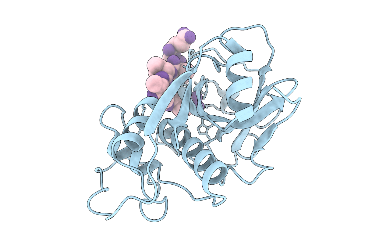
Title:
CRYSTAL STRUCTURE OF PAPAIN-SUCCINYL-GLN-VAL-VAL-ALA-ALA-P-NITROANILIDE COMPLEX AT 1.7 ANGSTROMS RESOLUTION: NONCOVALENT BINDING MODE OF A COMMON SEQUENCE OF ENDOGENOUS THIOL PROTEASE INHIBITORS
Biological Source:
Source Organism(s):
Carica papaya (Taxon ID: 3649)
Method Details:
Experimental Method:
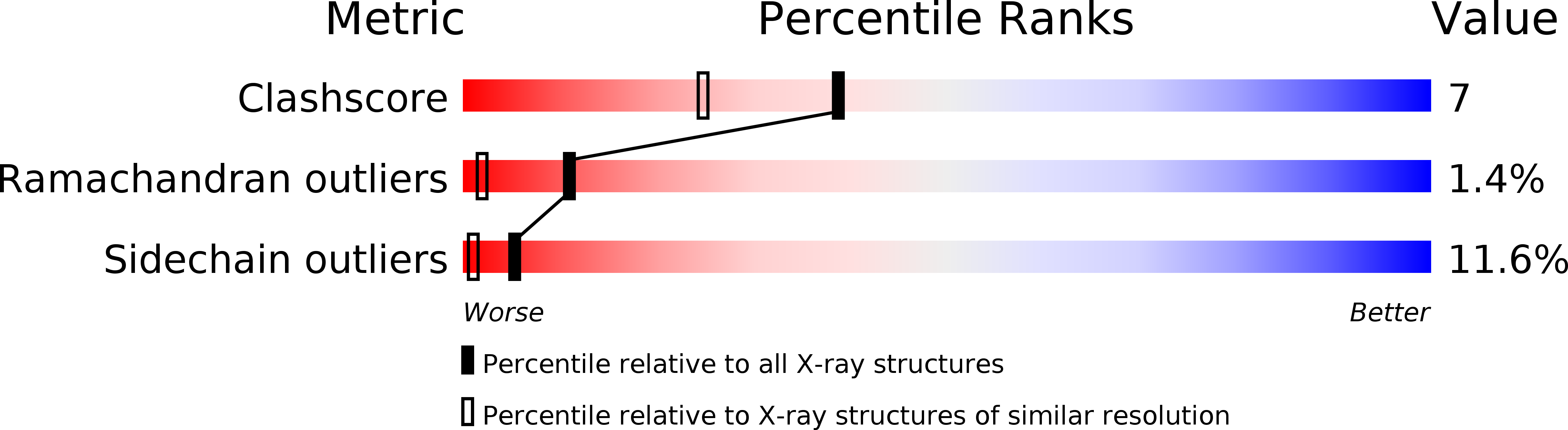
Resolution:
1.70 Å
R-Value Work:
0.19
R-Value Observed:
0.19
Space Group:
P 21 21 21