1n0u image
Deposition Date 2002-10-15
Release Date 2003-02-11
Last Version Date 2024-02-14
Entry Detail
PDB ID:
1N0U
Keywords:
Title:
Crystal structure of yeast elongation factor 2 in complex with sordarin
Biological Source:
Source Organism:
Method Details:
Experimental Method:
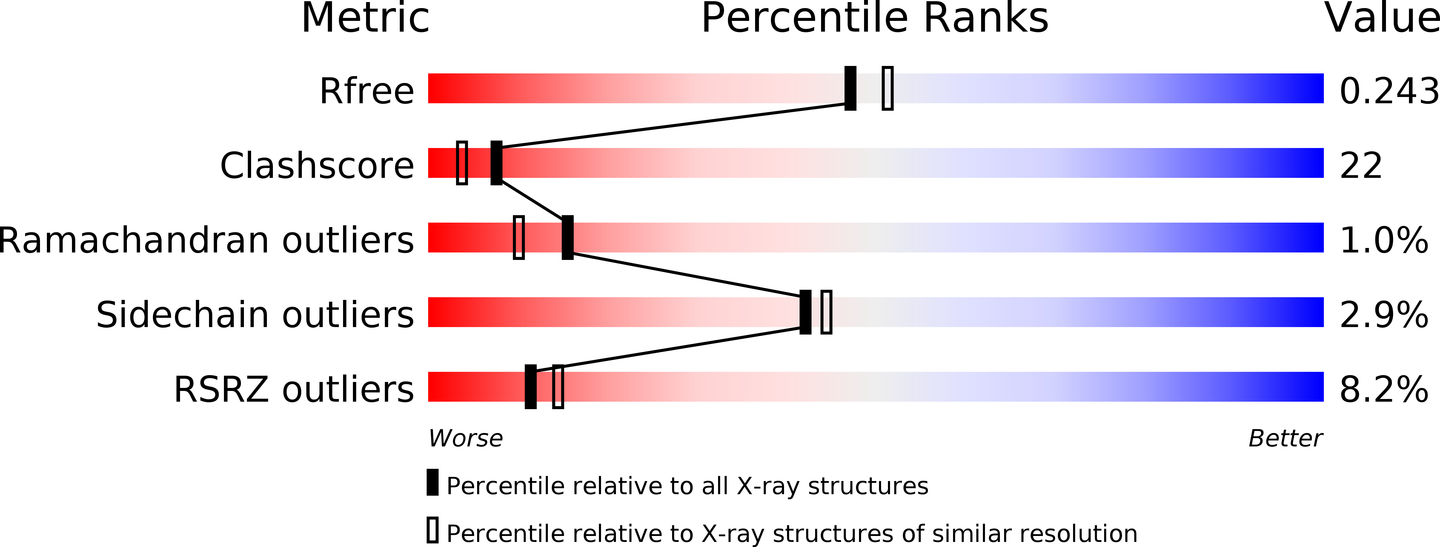
Resolution:
2.12 Å
R-Value Free:
0.25
R-Value Work:
0.23
R-Value Observed:
0.23
Space Group:
P 21 21 2
Macromolecular Entities
Structures with similar UniProt ID
Protein Blast
Polymer Type:polypeptide(L)
Molecule:Elongation factor 2
Gene (Uniprot):EFT1, EFT2
Chain IDs:A
Chain Length:842
Number of Molecules:1
Biological Source:Saccharomyces cerevisiae
Ligand Molecules
Primary Citation
Two crystal structures demonstrate large conformational changes in the eukaryotic ribosomal translocase.
Nat. Struct. Biol. 10 379 385 (2003)
PMID: 12692531 DOI: 10.1038/nsb923

Abstact

Two crystal structures of yeast translation elongation factor 2 (eEF2) were determined: the apo form at 2.9 A resolution and eEF2 in the presence of the translocation inhibitor sordarin at 2.1 A resolution. The overall conformation of apo eEF2 is similar to that of its prokaryotic homolog elongation factor G (EF-G) in complex with GDP. Upon sordarin binding, the three tRNA-mimicking C-terminal domains undergo substantial conformational changes, while the three N-terminal domains containing the nucleotide-binding site form an almost rigid unit. The conformation of eEF2 in complex with sordarin is entirely different from known conformations observed in crystal structures of EF-G or from cryo-EM studies of EF-G-70S complexes. The domain rearrangements induced by sordarin binding and the highly ordered drug-binding site observed in the eEF2-sordarin structure provide a high-resolution structural basis for the mechanism of sordarin inhibition. The two structures also emphasize the dynamic nature of the ribosomal translocase.

Legend

Protein

Chemical

Disease

Primary Citation of related structures