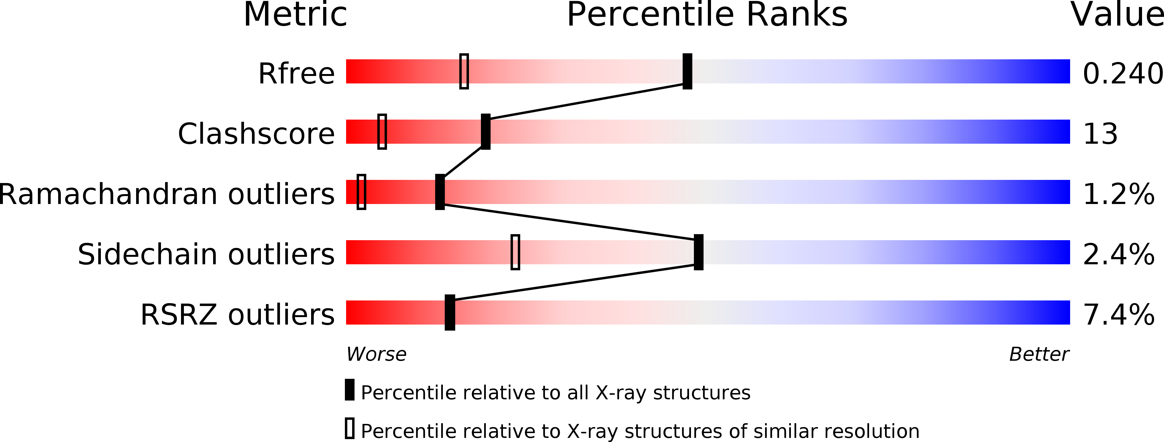
1mvf image
Deposition Date 2002-09-25
Release Date 2003-06-10
Last Version Date 2024-10-30
Entry Detail
PDB ID:
1MVF
Keywords:
Title:
MazE addiction antidote
Biological Source:
Source Organism:
Host Organism:
Method Details:
Experimental Method:
Resolution:
1.65 Å
R-Value Free:
0.24
R-Value Work:
0.21
R-Value Observed:
0.22
Space Group:
P 1 21 1
Macromolecular Entities
Polymer Type:polypeptide(L)
Molecule:immunoglobulin heavy chain variable region
Chain IDs:A, B
Chain Length:135
Number of Molecules:2
Biological Source:Camelus dromedarius
Polymer Type:polypeptide(L)
Molecule:PemI-like protein 1
Chain IDs:C (auth: D), D (auth: E)
Chain Length:82
Number of Molecules:2
Biological Source:Escherichia coli
Primary Citation
Crystal structure of the intrinsically flexible addiction antidote MazE.
J.Biol.Chem. 278 28252 28257 (2003)
PMID: 12743116 DOI: 10.1074/jbc.M302336200

Abstact

A specific camel VHH (variable domain of dromedary heavy chain antibody) fragment was used to crystallize the intrinsically flexible addiction antidote MazE. Only 45% of the polypeptide chain is found ordered in the crystal. The MazE monomer consisting of two beta-hairpins connected by a short alpha-helix has no hydrophobic core on its own and represents only one half of a typical protein domain. A complete domain structure is formed by the association of two chains, creating a hydrophobic core between two four-stranded beta-sheets. This hydrophobic core consists exclusively of short aliphatic residues. The folded part of MazE contains a novel DNA binding motif. A model for DNA binding that is consistent with the available biochemical data is presented.

Legend

Protein

Chemical

Disease

Primary Citation of related structures