1mgv image
Entry Detail
PDB ID:
1MGV
Keywords:
Title:
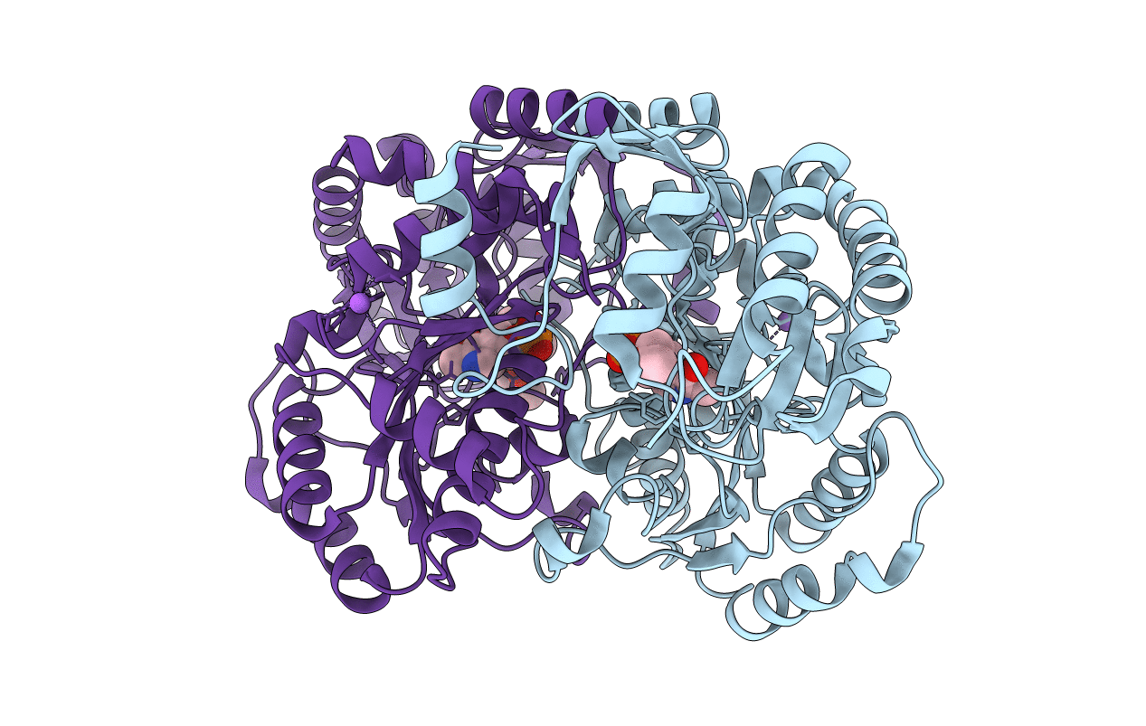
Crystal Structure of the R391A Mutant of 7,8-Diaminopelargonic Acid Synthase
Biological Source:
Source Organism:
PDB Version:
Deposition Date:
2002-08-16
Release Date:
2002-11-27
Method Details:
Experimental Method:
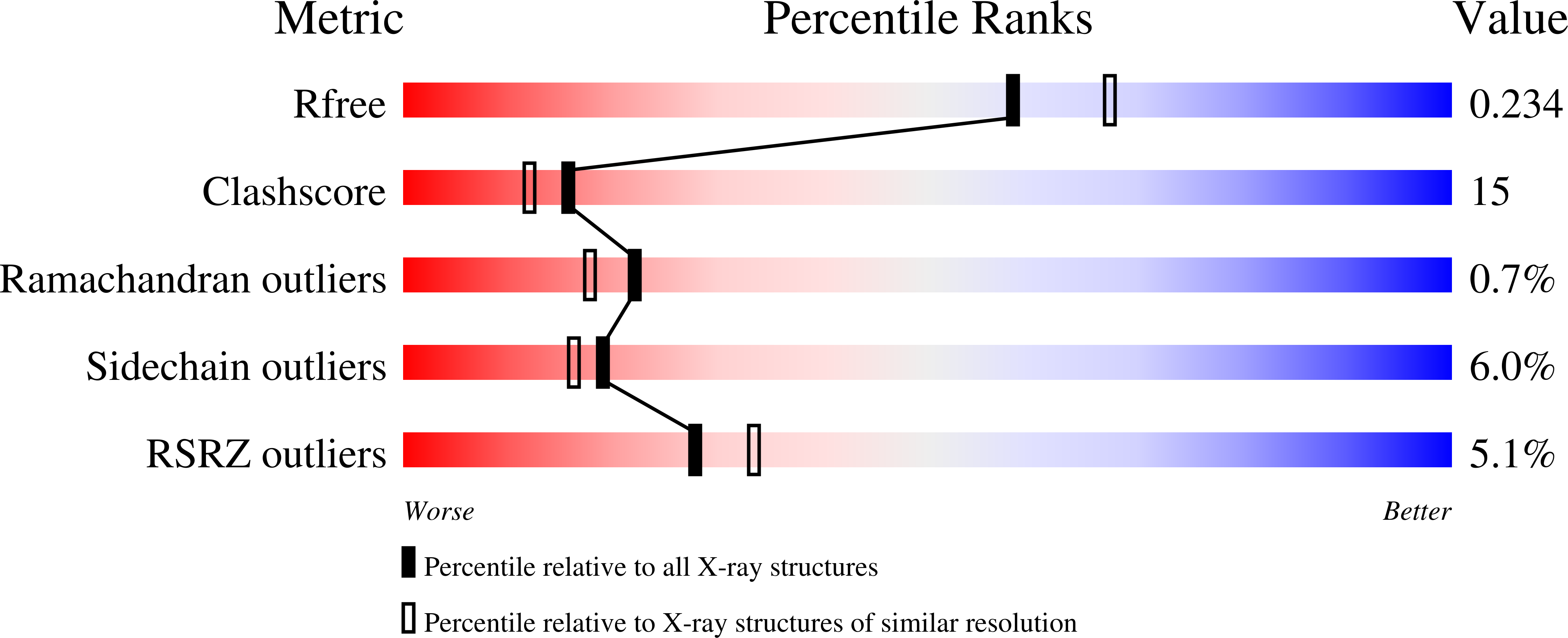
Resolution:
2.10 Å
R-Value Free:
0.23
R-Value Work:
0.20
R-Value Observed:
0.20
Space Group:
C 1 2 1
Macromolecular Entities
Polymer Type:polypeptide(L)
Description:7,8-diamino-pelargonic acid aminotransferase
Mutations:R391A
Chain IDs:A, B
Chain Length:429
Number of Molecules:2
Biological Source:Escherichia coli
Primary Citation
The Dual-Specific Active Site of 7,8-Diaminopelargonic Acid Synthase and the Effect of the R391A Mutation
Biochemistry 41 12582 12589 (2002)
PMID: 12379100 DOI: 10.1021/bi026339a

Abstact

7,8-diaminopelargonic acid (DAPA) synthase (EC 2.6.1.62) is a pyridoxal phosphate (PLP)-dependent transaminase that catalyzes the transfer of the alpha-amino group from S-adenosyl-L-methionine (SAM) to 7-keto-8-aminopelargonic acid (KAPA) to form DAPA in the antepenultimate step in the biosynthesis of biotin. The wild-type enzyme has a steady-state kcat value of 0.013 s(-1), and the K(m) values for SAM and KAPA are 150 and <2 microM, respectively. The k(max) and apparent K(m) values for the half-reaction of the PLP form of the enzyme with SAM are 0.016 s(-1) and 300 microM, respectively, while those for the reaction with DAPA are 0.79 s(-1) and 1 microM. The R391A mutant enzyme exhibits near wild-type kinetic parameters in the reaction with SAM, while the apparent K(m) for DAPA is increased 180-fold. The 2.1 A crystal structure of the R391A mutant enzyme shows that the mutation does not significantly alter the structure. These results indicate that the conserved arginine residue is not required for binding the alpha-amino acid SAM, but it is important for recognition of DAPA.

Legend

Protein

Chemical

Disease

Primary Citation of related structures