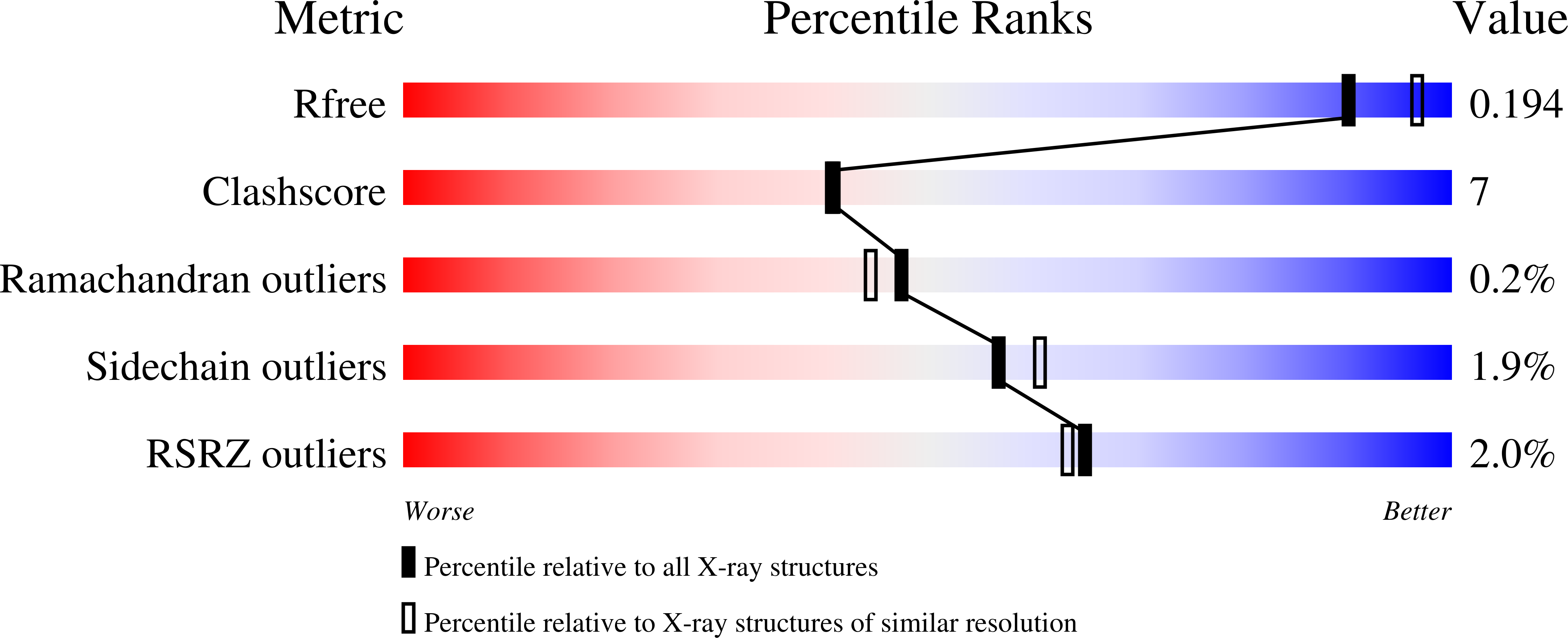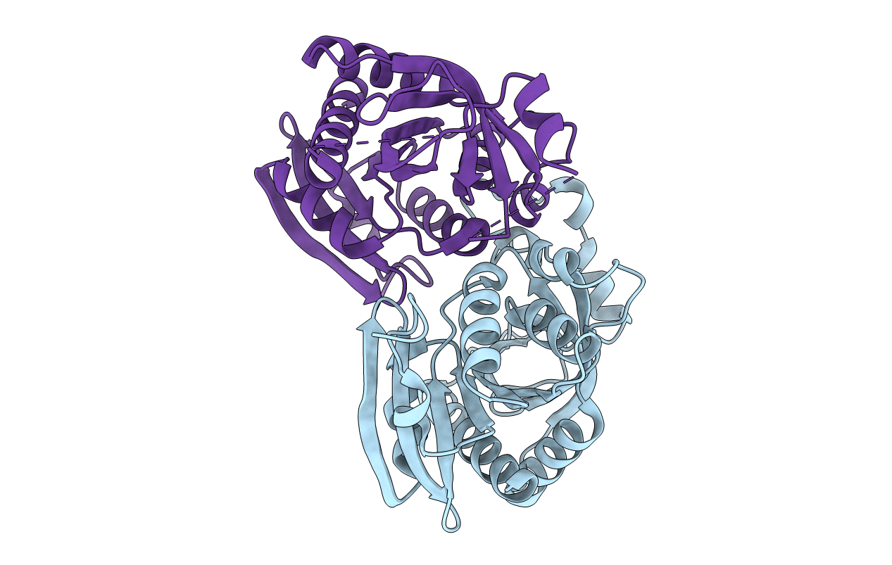
1lx7 image
Deposition Date 2002-06-04
Release Date 2002-06-12
Last Version Date 2024-10-30
Entry Detail
PDB ID:
1LX7
Keywords:
Title:
Structure of E. coli uridine phosphorylase at 2.0A
Biological Source:
Source Organism:
Escherichia coli (Taxon ID: 562)
Host Organism:
Method Details:
Experimental Method:
Resolution:
2.00 Å
R-Value Free:
0.21
R-Value Work:
0.18
Space Group:
H 3
Macromolecular Entities
Polymer Type:polypeptide(L)
Molecule:uridine phosphorylase
Gene (Uniprot):udp
Chain IDs:A, B
Chain Length:253
Number of Molecules:2
Biological Source:Escherichia coli
Modified Residue
Compound ID Chain ID Parent Comp ID Details 2D Image
MSE A MET SELENOMETHIONINE
Primary Citation
Structure of Escherichia coli uridine phosphorylase at 2.0 A.
Acta Crystallogr.,Sect.D 59 73 76 (2003)
PMID: 12499542 DOI: 10.1107/S0907444902018929

Abstact

The 2.0 A crystal structure has been determined for Escherichia coli uridine phosphorylase (UP), an essential enzyme in nucleotide biosynthesis that catalyzes the phosphorolytic cleavage of the C-N glycosidic bond of uridine to ribose-1-phosphate and uracil. The structure determination of two independent monomers in the asymmetric unit revealed the residue composition and atomic details of the apo configurations of each active site. The native hexameric UP enzyme was revealed by applying threefold crystallographic symmetry to the contents of the asymmetric unit. The 2.0 A model reveals a closer structural relationship to other nucleotide phosphorylase enzymes than was previously appreciated.

Legend

Protein

Chemical

Disease

Primary Citation of related structures