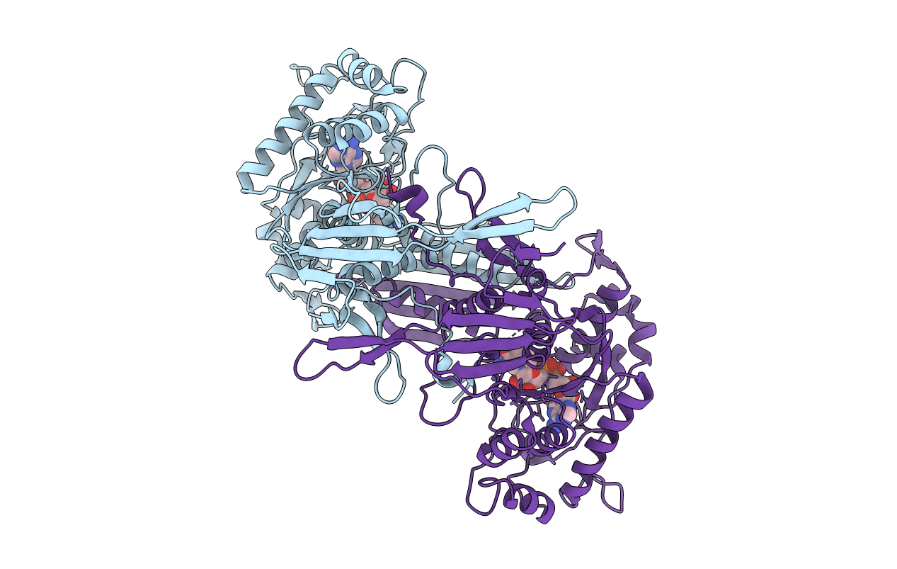
1jkf image
Deposition Date 2001-07-12
Release Date 2002-04-10
Last Version Date 2024-12-25
Entry Detail
PDB ID:
1JKF
Keywords:
Title:
Holo 1L-myo-inositol-1-phosphate Synthase
Biological Source:
Source Organism:
Host Organism:
Method Details:
Experimental Method:
Resolution:
2.40 Å
R-Value Free:
0.24
R-Value Work:
0.20
R-Value Observed:
0.24
Space Group:
C 1 2 1
Macromolecular Entities
Polymer Type:polypeptide(L)
Molecule:myo-inositol-1-phosphate synthase
Gene (Uniprot):INO1
Chain IDs:A, B
Chain Length:533
Number of Molecules:2
Biological Source:Saccharomyces cerevisiae
Ligand Molecules
Primary Citation
The crystal structure and mechanism of 1-L-myo-inositol- 1-phosphate synthase
J.Biol.Chem. 277 9484 9491 (2002)
PMID: 11779862 DOI: 10.1074/jbc.M109371200

Abstact

1-l-myo-Inositol-1-phosphate synthase catalyzes the conversion of d-glucose 6-phosphate to 1-l-myo-inositol-1-phosphate (MIP), the first and rate-limiting step in the biosynthesis of all inositol-containing compounds. It involves an oxidation, intramolecular aldol cyclization, and reduction. We have determined the first crystal structure of MIP synthase. We present structures of both the NAD-bound enzyme and the enzyme bound to an inhibitor, 2-deoxy-glucitol-6-phosphate. While 58 amino acids are disordered in the unbound form of the enzyme in the vicinity of the active site, the inhibitor nucleates the folding of this domain in a striking example of induced fit, serving to completely encapsulate it within the enzyme. Three helices and a long beta-strand are formed in this process. We postulate a mechanism for the conversion based on the structure of the inhibitor-bound complex.

Legend

Protein

Chemical

Disease

Primary Citation of related structures