Deposition Date
2002-04-25
Release Date
2003-12-23
Last Version Date
2024-10-30
Entry Detail
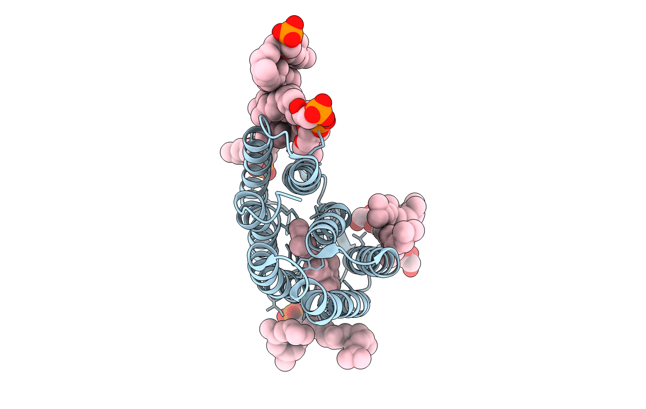
PDB ID:
1IW9
Keywords:
Title:
Crystal Structure of the M Intermediate of Bacteriorhodopsin
Biological Source:
Source Organism(s):
Halobacterium salinarum (Taxon ID: 2242)
Method Details:
Experimental Method:
Resolution:
2.50 Å
R-Value Free:
0.27
R-Value Work:
0.22
R-Value Observed:
0.22
Space Group:
P 6 2 2