1iat image
Deposition Date 2001-03-23
Release Date 2001-05-30
Last Version Date 2024-04-03
Entry Detail
PDB ID:
1IAT
Keywords:
Title:
CRYSTAL STRUCTURE OF HUMAN PHOSPHOGLUCOSE ISOMERASE/NEUROLEUKIN/AUTOCRINE MOTILITY FACTOR/MATURATION FACTOR
Biological Source:
Source Organism:
Homo sapiens (Taxon ID: 9606)
Host Organism:
Method Details:
Experimental Method:
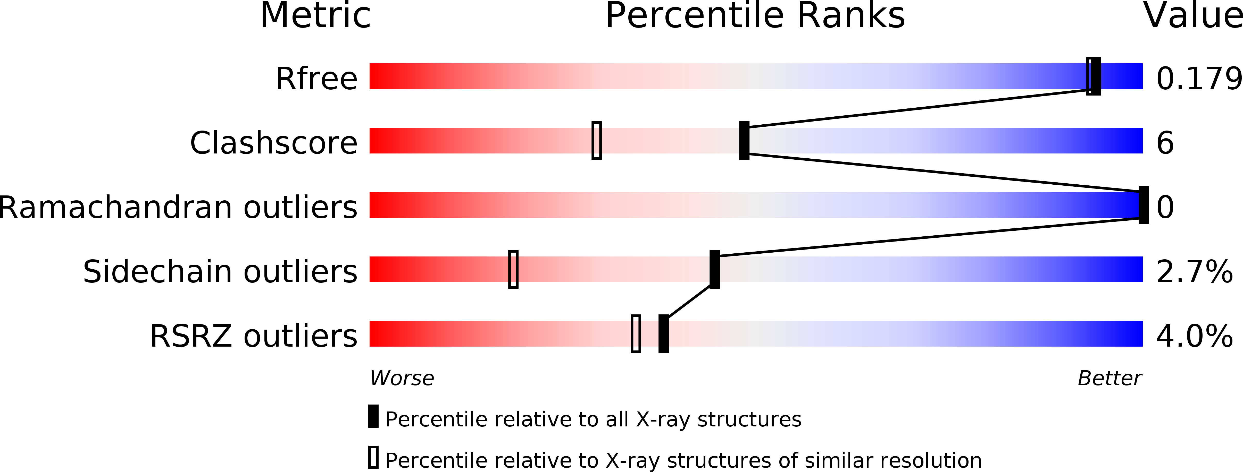
Resolution:
1.62 Å
R-Value Free:
0.17
R-Value Work:
0.14
R-Value Observed:
0.14
Space Group:
P 43 21 2
Macromolecular Entities
Polymer Type:polypeptide(L)
Molecule:PHOSPHOGLUCOSE ISOMERASE
Gene (Uniprot):GPI
Chain IDs:A
Chain Length:557
Number of Molecules:1
Biological Source:Homo sapiens
Primary Citation
The crystal structure of human phosphoglucose isomerase at 1.6 A resolution: implications for catalytic mechanism, cytokine activity and haemolytic anaemia.
J.Mol.Biol. 309 447 463 (2001)
PMID: 11371164 DOI: 10.1006/jmbi.2001.4680

Abstact

Phosphoglucose isomerase (PGI) is a multifunctional protein, which, inside the cell, functions as a housekeeping enzyme of glycolysis and gluconeogenesis and, outside the cell, exerts wholly unrelated cytokine properties. We have determined the structure of human PGI to a resolution of 1.6 A using X-ray crystallography. The structure is highly similar to other PGIs, especially the architecture of the active site. Fortuitous binding of a sulphate molecule from the crystallisation solution has facilitated an accurate description of the substrate phosphate-binding site. Comparison with both native and inhibitor-bound rabbit PGI structures shows that two loops move closer to the active site upon binding inhibitor. Interestingly, the human structure most closely resembles the inhibitor-bound structure, suggesting that binding of the phosphate moiety of the substrate may trigger this conformational change. We suggest a new mechanism for catalysis that uses Glu357 as the base catalyst for the isomerase reaction rather than His388 as proposed previously. The human PGI structure has also provided a detailed framework with which to map mutations associated with non-spherocytic haemolytic anaemia.

Legend

Protein

Chemical

Disease

Primary Citation of related structures