1hox image
Deposition Date 2000-12-11
Release Date 2001-07-20
Last Version Date 2023-08-09
Entry Detail
PDB ID:
1HOX
Keywords:
Title:
CRYSTAL STRUCTURE OF RABBIT PHOSPHOGLUCOSE ISOMERASE COMPLEXED WITH FRUCTOSE-6-PHOSPHATE
Biological Source:
Source Organism:
Method Details:
Experimental Method:
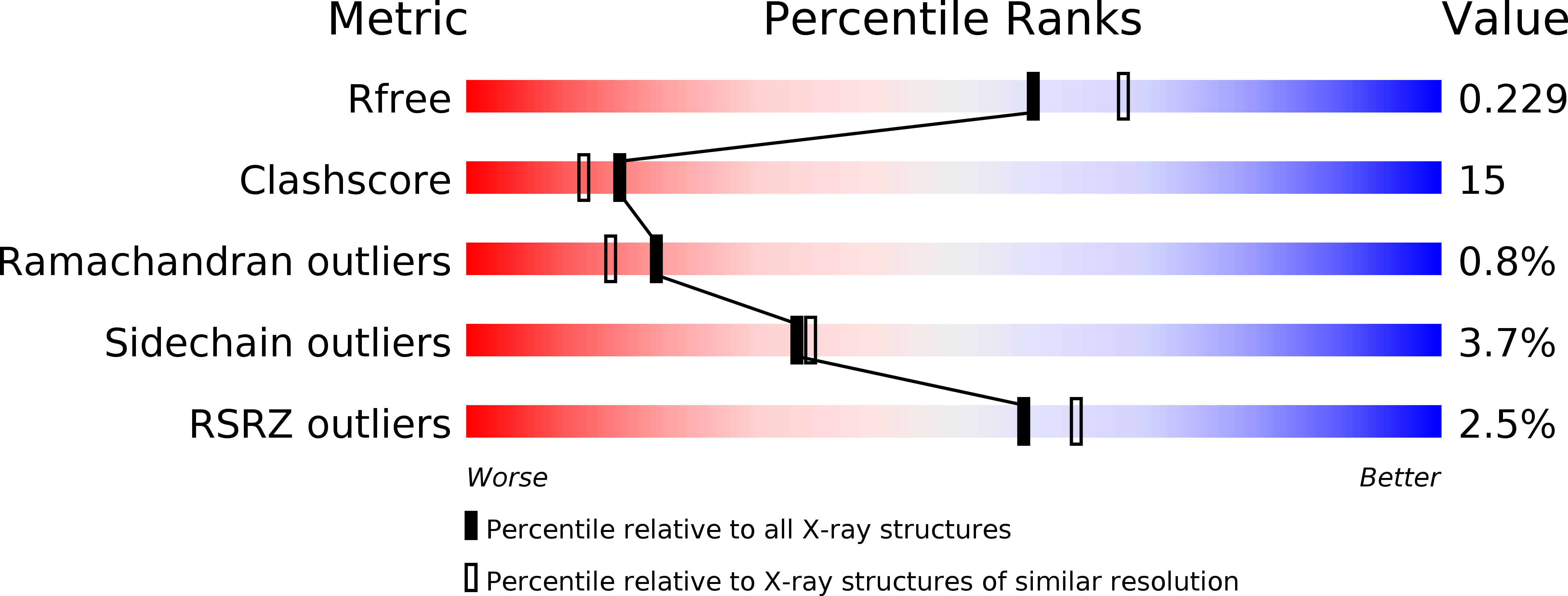
Resolution:
2.10 Å
R-Value Free:
0.24
R-Value Work:
0.21
R-Value Observed:
0.22
Space Group:
C 2 2 21
Macromolecular Entities
Polymer Type:polypeptide(L)
Molecule:PHOSPHOGLUCOSE ISOMERASE
Gene (Uniprot):GPI
Chain IDs:A, B
Chain Length:558
Number of Molecules:2
Biological Source:Oryctolagus cuniculus
Ligand Molecules
Primary Citation
Crystal structure of rabbit phosphoglucose isomerase complexed with its substrate D-fructose 6-phosphate.
Biochemistry 40 7799 7805 (2001)
PMID: 11425306 DOI: 10.1021/bi002916o

Abstact

Phosphoglucose isomerase (PGI, EC 5.3.1.9) catalyzes the interconversion of D-glucose 6-phosphate (G6P) and D-fructose 6-phosphate (F6P) and plays important roles in glycolysis and gluconeogenesis. Biochemical characterization of the enzyme has led to a proposed multistep catalytic mechanism. First, the enzyme catalyzes ring opening to yield the open chain form of the substrate. Then isomerization proceeds via proton transfer between C2 and C1 of a cis-enediol(ate) intermediate to yield the open chain form of the product. Catalysis proceeds in both the G6P to F6P and F6P to G6P directions, so both G6P and F6P are substrates. X-ray crystal structure analysis of rabbit and bacterial PGI has previously identified the location of the enzyme active site, and a recent crystal structure of rabbit PGI identified Glu357 as a candidate functional group for transferring the proton. However, it was not clear which active site amino acid residues catalyze the ring opening step. In this paper, we report the X-ray crystal structure of rabbit PGI complexed with the cyclic form of its substrate, D-fructose 6-phosphate, at 2.1 A resolution. The location of the substrate relative to the side chains of His388 suggest that His388 promotes ring opening by protonating the ring oxygen. Glu216 helps to position His388, and a water molecule that is held in position by Lys518 and Thr214 accepts a proton from the hydroxyl group at C2. Comparison to a structure of rabbit PGI with 5PAA bound indicates that ring opening is followed by loss of the protonated water molecule and conformational changes in the substrate and the protein so that a helix containing amino acids 513-520 moves in toward the substrate to form additional hydrogen bonds with the substrate.

Legend

Protein

Chemical

Disease

Primary Citation of related structures