Deposition Date
2001-12-10
Release Date
2002-03-12
Last Version Date
2024-05-01
Entry Detail
PDB ID:
1GR0
Keywords:
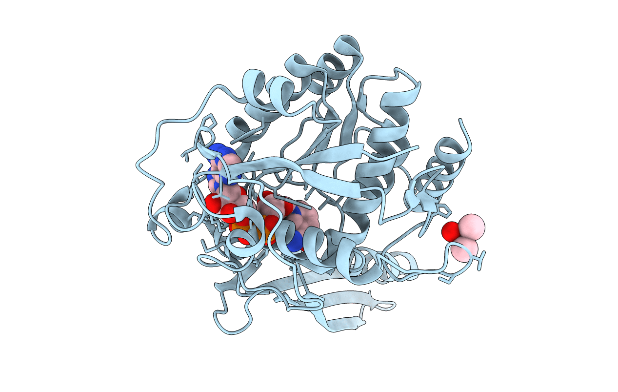
Title:
myo-inositol 1-phosphate synthase from Mycobacterium tuberculosis in complex with NAD and zinc.
Biological Source:
Source Organism(s):
MYCOBACTERIUM TUBERCULOSIS (Taxon ID: 1773)
Expression System(s):
Method Details:
Experimental Method:
Resolution:
1.95 Å
R-Value Free:
0.23
R-Value Work:
0.21
R-Value Observed:
0.21
Space Group:
P 4 21 2