1fz2 image
Deposition Date 2000-10-03
Release Date 2001-02-21
Last Version Date 2024-02-07
Entry Detail
PDB ID:
1FZ2
Keywords:
Title:
METHANE MONOOXYGENASE HYDROXYLASE, FORM II MIXED-VALENT GENERATED BY CRYSTAL SOAKING
Biological Source:
Source Organism:
Method Details:
Experimental Method:
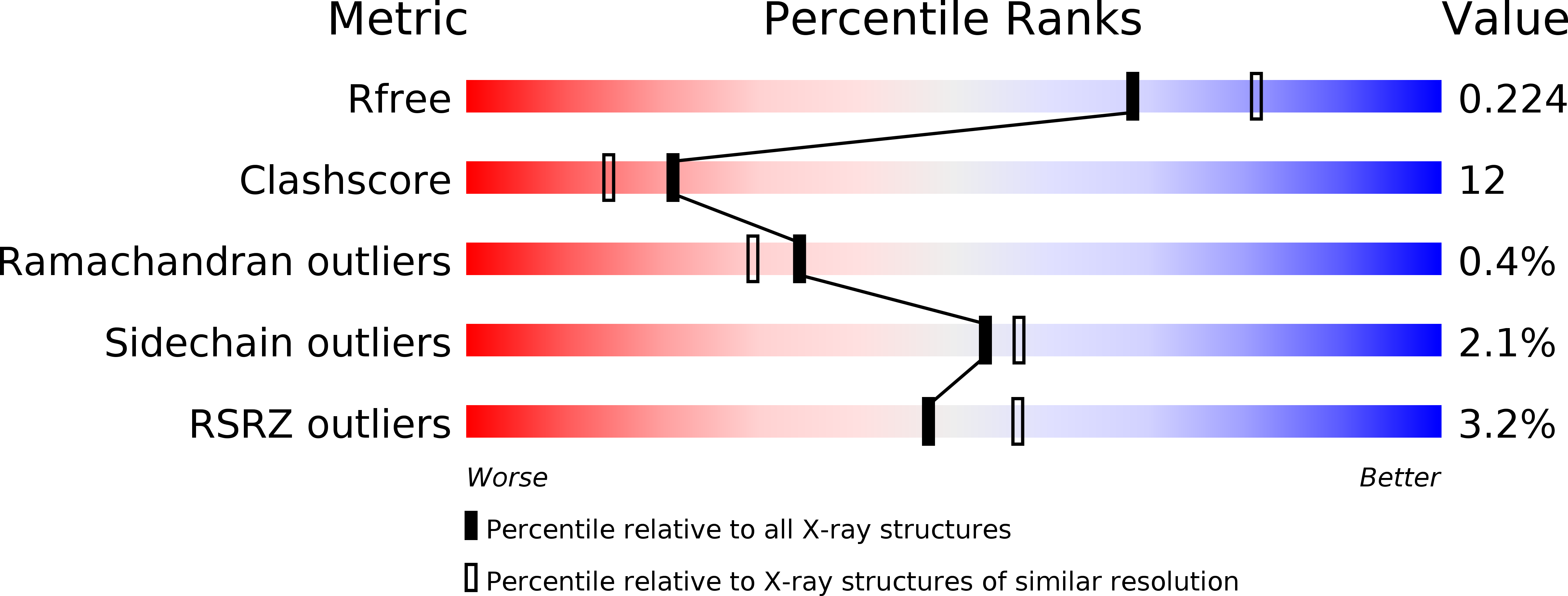
Resolution:
2.15 Å
R-Value Free:
0.22
R-Value Work:
0.19
R-Value Observed:
0.19
Space Group:
P 21 21 21
Macromolecular Entities
Structures with similar UniProt ID
Protein Blast
Polymer Type:polypeptide(L)
Molecule:METHANE MONOOXYGENASE COMPONENT A, ALPHA CHAIN
Gene (Uniprot):mmoX
Chain IDs:A, B
Chain Length:527
Number of Molecules:2
Biological Source:Methylococcus capsulatus
Structures with similar UniProt ID
Protein Blast
Polymer Type:polypeptide(L)
Molecule:METHANE MONOOXYGENASE COMPONENT A, BETA CHAIN
Gene (Uniprot):mmoY
Chain IDs:C, D
Chain Length:389
Number of Molecules:2
Biological Source:Methylococcus capsulatus
Structures with similar UniProt ID
Protein Blast
Polymer Type:polypeptide(L)
Molecule:METHANE MONOOXYGENASE COMPONENT A, GAMMA CHAIN
Gene (Uniprot):mmoZ
Chain IDs:E, F
Chain Length:170
Number of Molecules:2
Biological Source:Methylococcus capsulatus
Primary Citation
Crystal structures of the soluble methane monooxygenase hydroxylase from Methylococcus capsulatus (Bath) demonstrating geometrical variability at the dinuclear iron active site.
J.Am.Chem.Soc. 123 827 838 (2001)
PMID: 11456616 DOI: 10.1021/ja003240n

Abstact

The oxidation of methane to methanol is performed at carboxylate-bridged dinuclear iron centers in the soluble methane monooxygenase hydroxylase (MMOH). Previous structural studies of MMOH, and the related R2 subunit of ribonucleotide reductase, have demonstrated the occurrence of carboxylate shifts involving glutamate residues that ligate the catalytic iron atoms. These shifts are thought to have important mechanistic implications. Recent kinetic and theoretical studies have also emphasized the importance of hydrogen bonding and pH effects at the active site. We report here crystal structures of MMOH from Methylococcus capsulatus (Bath) in the diiron(II), diiron(III), and mixed-valent Fe(II)Fe(III) oxidation states, and at pH values of 6.2, 7.0, and 8.5. These structures were investigated in an effort to delineate the range of possible motions at the MMOH active site and to identify hydrogen-bonding interactions that may be important in understanding catalysis by the enzyme. Our results present the first view of the diiron center in the mixed-valent state, and they indicate an increased lability for ferrous ions in the enzyme. Alternate conformations of Asn214 near the active site according to redox state and a distortion in one of the alpha-helices adjacent to the metal center in the diiron(II) state have also been identified. These changes alter the surface of the protein in the vicinity of the catalytic core and may have implications for small-molecule accessibility to the active site and for protein component interactions in the methane monooxygenase system. Collectively, these results help to explain previous spectroscopic observations and provide new insight into catalysis by the enzyme.

Legend

Protein

Chemical

Disease

Primary Citation of related structures
Feedback Form
Name
Email
Institute
Feedback