Deposition Date
1993-03-04
Release Date
1994-01-31
Last Version Date
2024-10-23
Entry Detail
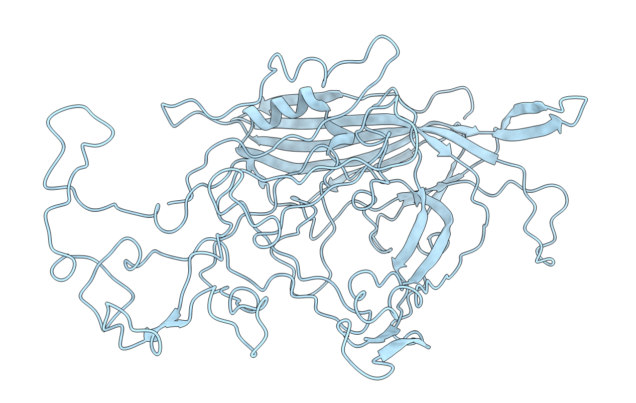
PDB ID:
1FPV
Keywords:
Title:
STRUCTURE DETERMINATION OF FELINE PANLEUKOPENIA VIRUS EMPTY PARTICLES
Biological Source:
Source Organism(s):
Feline panleukopenia virus (Taxon ID: 10786)
Method Details: