Deposition Date
2000-07-17
Release Date
2001-07-25
Last Version Date
2024-10-16
Entry Detail
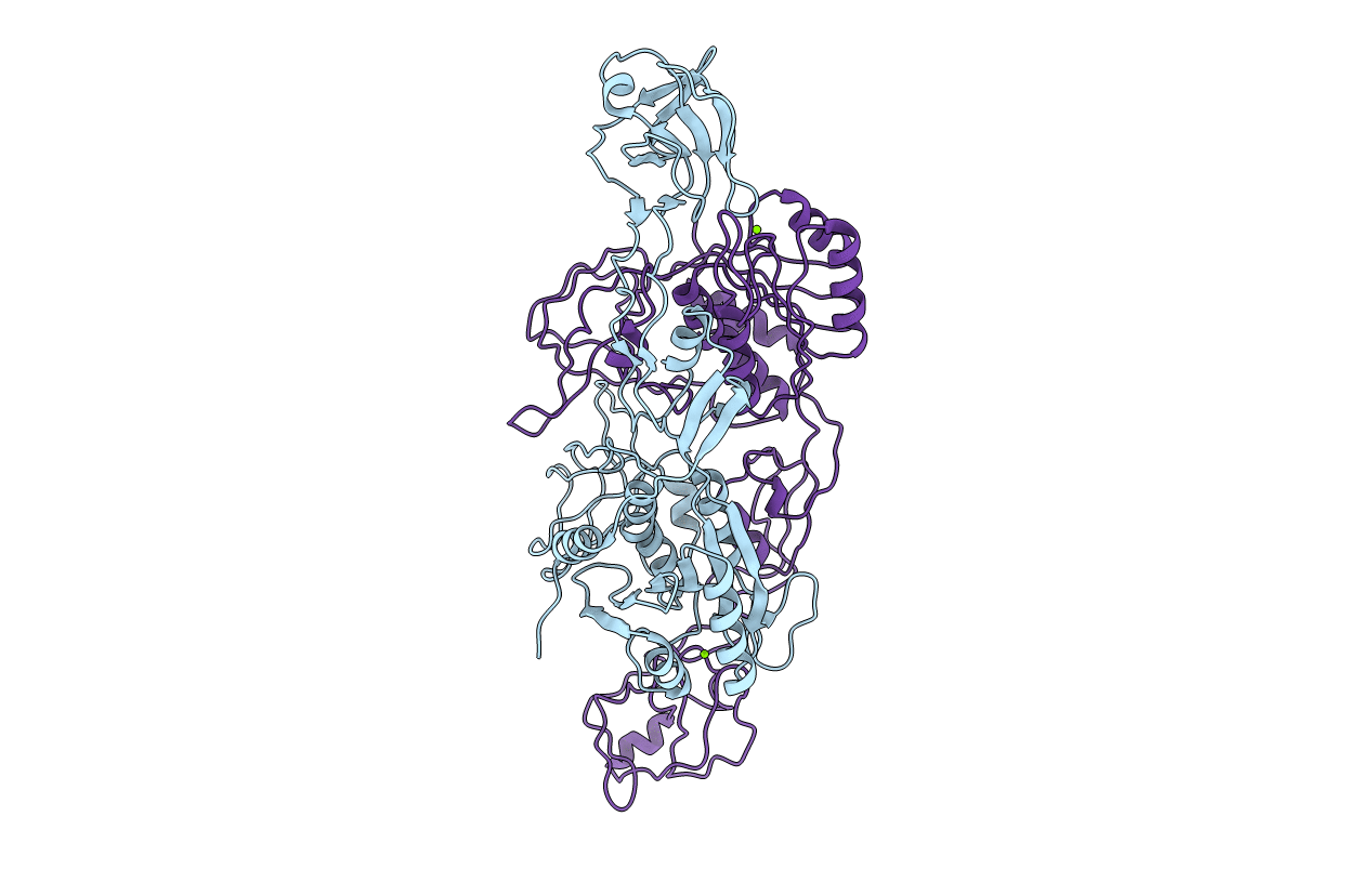
PDB ID:
1FC5
Keywords:
Title:
CRYSTAL STRUCTURE OF MOLYBDOPTERIN BIOSYNTHESIS MOEA PROTEIN
Biological Source:
Source Organism(s):
Escherichia coli (Taxon ID: 562)
Expression System(s):
Method Details:
Experimental Method:
Resolution:
2.20 Å
R-Value Free:
0.29
R-Value Work:
0.25
R-Value Observed:
0.26
Space Group:
P 21 21 21