
Deposition Date
1981-05-21
Release Date
1981-10-02
Last Version Date
2024-11-20
Entry Detail
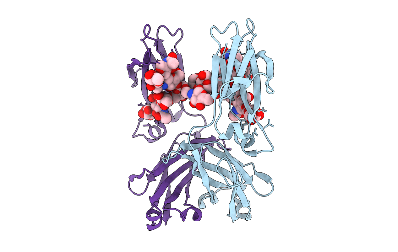
PDB ID:
1FC1
Keywords:
Title:
CRYSTALLOGRAPHIC REFINEMENT AND ATOMIC MODELS OF A HUMAN FC FRAGMENT AND ITS COMPLEX WITH FRAGMENT B OF PROTEIN A FROM STAPHYLOCOCCUS AUREUS AT 2.9-AND 2.8-ANGSTROMS RESOLUTION
Biological Source:
Source Organism(s):
Homo sapiens (Taxon ID: 9606)
Method Details:
