Deposition Date
2000-07-05
Release Date
2001-01-17
Last Version Date
2024-11-20
Entry Detail
PDB ID:
1F8U
Keywords:
Title:
CRYSTAL STRUCTURE OF MUTANT E202Q OF HUMAN ACETYLCHOLINESTERASE COMPLEXED WITH GREEN MAMBA VENOM PEPTIDE FASCICULIN-II
Biological Source:
Source Organism(s):
Homo sapiens (Taxon ID: 9606)
Dendroaspis angusticeps (Taxon ID: 8618)
Dendroaspis angusticeps (Taxon ID: 8618)
Expression System(s):
Method Details:
Experimental Method:
Resolution:
2.90 Å
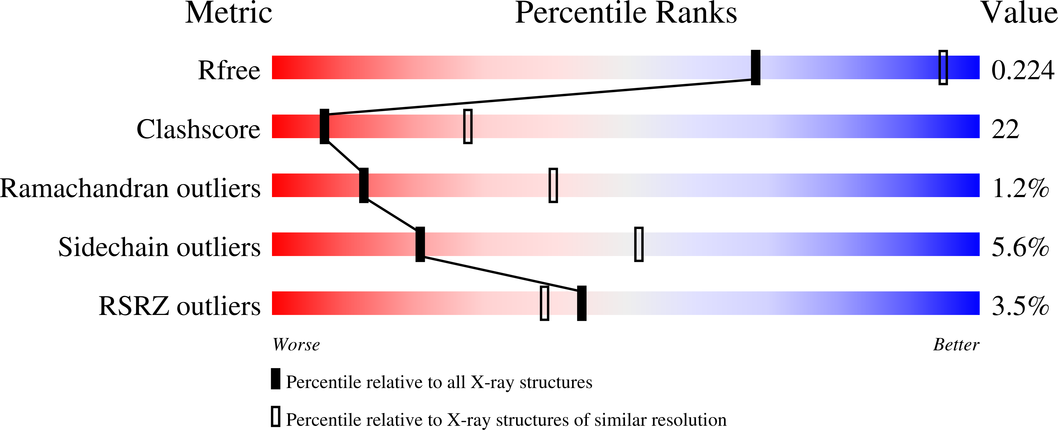
R-Value Free:
0.22
R-Value Work:
0.19
R-Value Observed:
0.19
Space Group:
H 3 2