1dvp image
Entry Detail
PDB ID:
1DVP
Keywords:
Title:
CRYSTAL STRUCTURE OF THE VHS AND FYVE TANDEM DOMAINS OF HRS, A PROTEIN INVOLVED IN MEMBRANE TRAFFICKING AND SIGNAL TRANSDUCTION
Biological Source:
Source Organism:
Host Organism:
PDB Version:
Deposition Date:
2000-01-21
Release Date:
2000-03-06
Method Details:
Experimental Method:
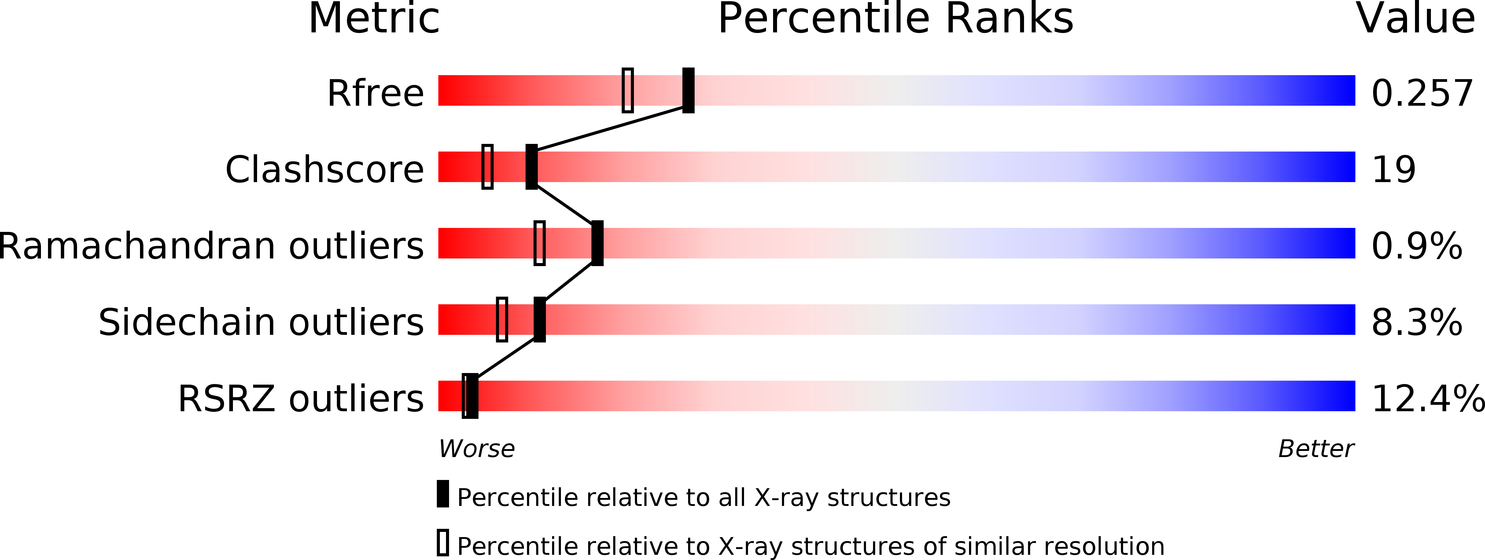
Resolution:
2.00 Å
R-Value Free:
0.25
R-Value Work:
0.21
R-Value Observed:
0.21
Space Group:
C 1 2 1
Macromolecular Entities
Polymer Type:polypeptide(L)
Description:HEPATOCYTE GROWTH FACTOR-REGULATED TYROSINE KINASE SUBSTRATE
Chain IDs:A
Chain Length:220
Number of Molecules:1
Biological Source:Drosophila melanogaster
Primary Citation
Crystal structure of the VHS and FYVE tandem domains of Hrs, a protein involved in membrane trafficking and signal transduction.
Cell(Cambridge,Mass.) 100 447 456 (2000)
PMID: 10693761 DOI: 10.1016/S0092-8674(00)80680-7

Abstact

We have determined the 2 A X-ray structure of the 219-residue N-terminal VHS and FYVE tandem domain unit of Drosophila Hrs. The unit assumes a pyramidal structure in which the much larger VHS domain (residues 1-153) forms a rectangular base and the FYVE domain occupies the apical end. The VHS domain is comprised of an unusual "superhelix" of eight alpha helices, and the FYVE domain is mainly built of loops, two double-stranded antiparallel sheets, and a helix stabilized by two tetrahedrally coordinated zinc atoms. The two-domain structure forms an exact 2-fold-related homodimer through antiparallel association of mainly FYVE domains. Dimerization creates two identical pockets designed for binding ligands with multiple negative charges such as citrate or phosphatidylinositol 3-phosphate.

Legend

Protein

Chemical

Disease

Primary Citation of related structures