1dgl image
Deposition Date 1998-02-10
Release Date 1999-06-01
Last Version Date 2024-05-22
Entry Detail
PDB ID:
1DGL
Keywords:
Title:
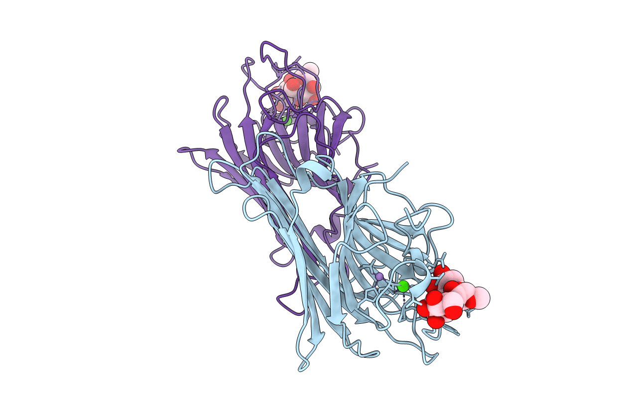
LECTIN FROM DIOCLEA GRANDIFLORA COMPLEXED TO TRIMANNOSIDE
Biological Source:
Source Organism:
Method Details:
Experimental Method:
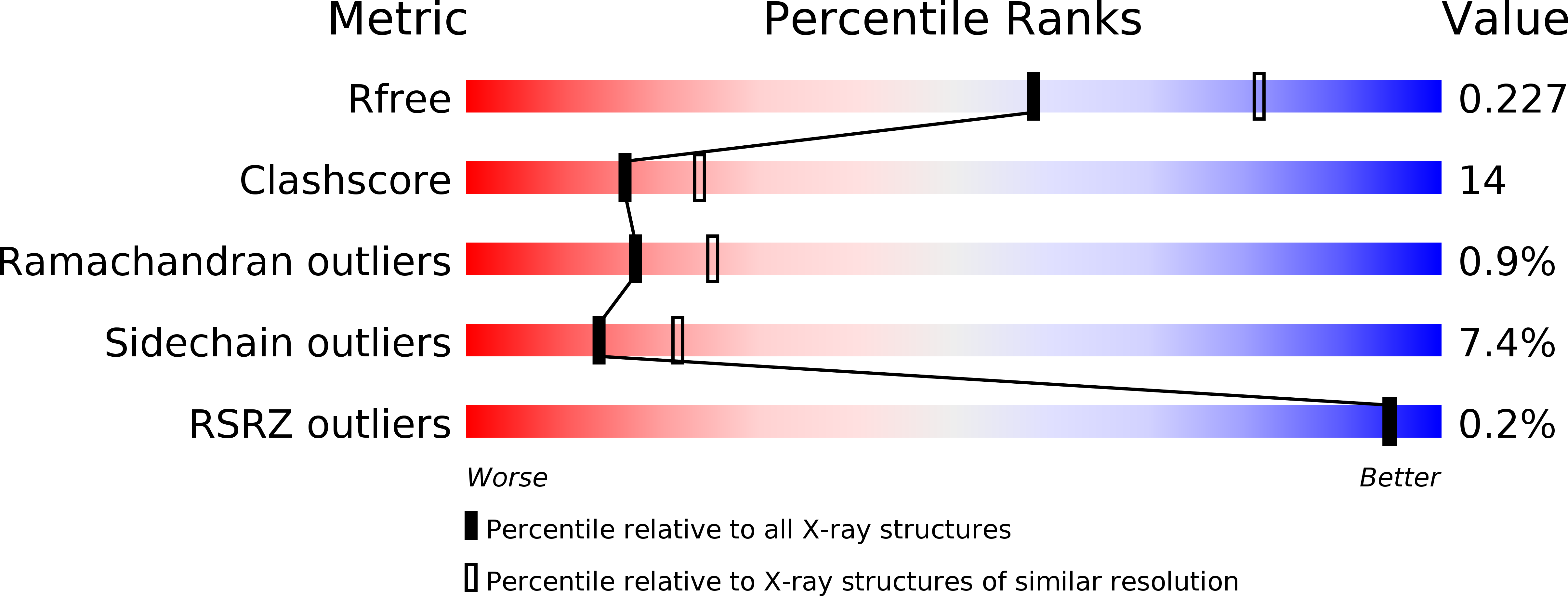
Resolution:
2.40 Å
R-Value Free:
0.25
R-Value Work:
0.18
R-Value Observed:
0.18
Space Group:
P 31 2 1
Macromolecular Entities
Polymer Type:polypeptide(L)
Molecule:LECTIN
Chain IDs:A, B
Chain Length:237
Number of Molecules:2
Biological Source:Dioclea grandiflora
Primary Citation
Crystal structure of the lectin from Dioclea grandiflora complexed with core trimannoside of asparagine-linked carbohydrates.
J.Biol.Chem. 273 32818 32825 (1998)
PMID: 9830028 DOI: 10.1074/jbc.273.49.32818

Abstact

The seed lectin from Dioclea grandiflora (DGL) has recently been shown to possess high affinity for 3, 6-di-O-(alpha-D-mannopyranosyl)-alpha-D-mannopyranose, the core trimannoside of asparagine-linked carbohydrates, but lower affinity for biantennary complex carbohydrates. In the previous paper, the thermodynamics of DGL binding to deoxy analogs of the core trimannoside and to a biantennary complex carbohydrate were determined by isothermal titration microcalorimetry. The data suggest that DGL recognizes specific hydroxyl groups of the trimannoside similar to that of the jack bean lectin concanavalin A (ConA) (Gupta, D. Dam, T. K., Oscarson, S., and Brewer, C. F. (1997) J. Biol. Chem. 272, 6388-6392). However, the thermodynamics of DGL binding to certain deoxy analogs and to the complex carbohydrate are different from that of ConA. In the present paper, the x-ray crystal structure of DGL complexed to the core trimannoside was determined to a resolution of 2.6 A. The overall structure of the DGL complex is similar to the structure of the ConA-trimannoside complex (Naismith, J. H., and Field, R. A. (1996) J. Biol. Chem. 271, 972-976). The location and conformation of the bound trimannoside as well as its hydrogen-bonding interactions in both complexes are nearly identical. However, differences exist in the location of two loops outside of the respective binding sites containing residues 114-125 and 222-227. The latter residues affect the location of a network of hydrogen-bonded water molecules that interact with the trisaccharide. Differences in the arrangement of ordered water molecules in the binding site and/or protein conformational differences outside of the binding site may account for the differences in the thermodynamics of binding of the two lectins to deoxy analogs of the trimannoside. Molecular modeling studies suggest how DGL discriminates against binding the biantennary complex carbohydrate relative to ConA.

Legend

Protein

Chemical

Disease

Primary Citation of related structures