
Deposition Date
1999-10-06
Release Date
1999-10-14
Last Version Date
2024-02-07
Entry Detail
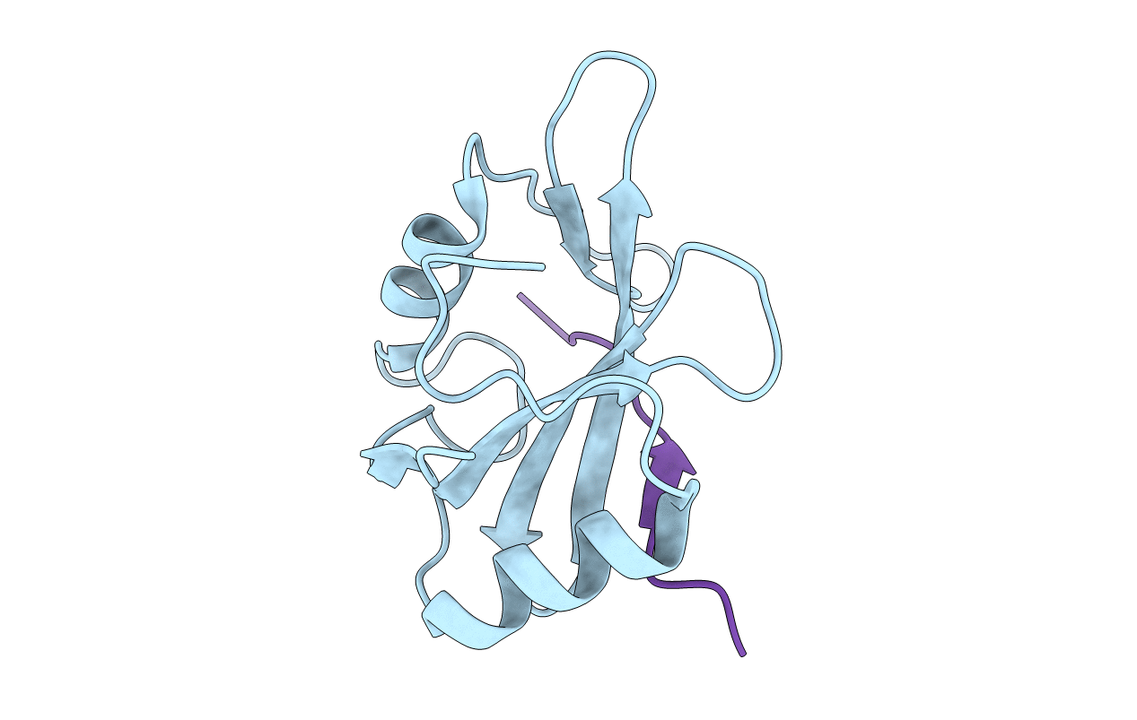
PDB ID:
1D4T
Keywords:
Title:
CRYSTAL STRUCTURE OF THE XLP PROTEIN SAP IN COMPLEX WITH A SLAM PEPTIDE
Biological Source:
Source Organism(s):
Homo sapiens (Taxon ID: 9606)
Expression System(s):
Method Details:
Experimental Method:
Resolution:
1.10 Å
R-Value Free:
0.16
R-Value Work:
0.11
R-Value Observed:
0.13
Space Group:
P 1 21 1
