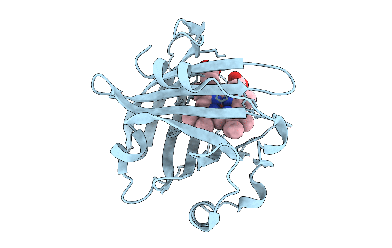
1d3s image
Deposition Date 1999-09-30
Release Date 2000-07-07
Last Version Date 2024-10-30
Entry Detail
PDB ID:
1D3S
Title:
1.4 A crystal structure of nitrophorin 4 from Rhodnius prolixis at pH=5.6.
Biological Source:
Source Organism:
Method Details:
Experimental Method:
Resolution:
1.40 Å
R-Value Free:
0.25
R-Value Work:
0.21
R-Value Observed:
0.21
Space Group:
C 1 2 1
Macromolecular Entities
Polymer Type:polypeptide(L)
Molecule:NITROPHORIN 4
Chain IDs:A
Chain Length:184
Number of Molecules:1
Biological Source:Rhodnius prolixus
Ligand Molecules
Primary Citation
Nitric oxide binding to nitrophorin 4 induces complete distal pocket burial.
Nat.Struct.Biol. 7 551 554 (2000)
PMID: 10876239 DOI: 10.1038/76769

Abstact

The nitrophorins comprise an unusual family of proteins that use ferric (Fe(III)) heme to transport highly reactive nitric oxide (NO) from the salivary gland of a blood sucking bug to the victim, resulting in vasodilation and reduced blood coagulation. We have determined structures of nitrophorin 4 in complexes with H2O, cyanide and nitric oxide. These structures reveal a remarkable feature: the nitrophorins have a broadly open distal pocket in the absence of NO, but upon NO binding, three or more water molecules are expelled and two loops fold into the distal pocket, resulting in the packing of hydrophobic groups around the NO molecule and increased distortion of the heme. In this way, the protein apparently forms a 'hydrophobic trap' for the NO molecule. The structures are very accurate, ranging between 1.6 and 1.4 A resolutions.

Legend

Protein

Chemical

Disease

Primary Citation of related structures