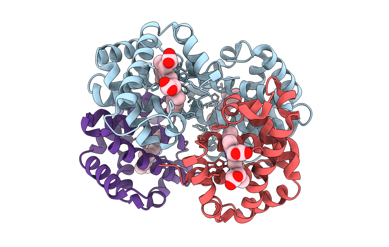
1c7d image
Deposition Date 2000-02-09
Release Date 2000-06-30
Last Version Date 2023-08-09
Entry Detail
PDB ID:
1C7D
Title:
DEOXY RHB1.2 (RECOMBINANT HEMOGLOBIN)
Biological Source:
Source Organism:
Homo sapiens (Taxon ID: 9606)
Host Organism:
Method Details:
Experimental Method:
Resolution:
1.80 Å
R-Value Free:
0.23
R-Value Observed:
0.18
Space Group:
P 1 21 1
Macromolecular Entities
Polymer Type:polypeptide(L)
Molecule:PROTEIN (DEOXYHEMOGLOBIN (ALPHA CHAIN))
Gene (Uniprot):HBA1, HBA2
Mutations:V1M,142G,143G
Chain IDs:A
Chain Length:284
Number of Molecules:1
Biological Source:Homo sapiens
Polymer Type:polypeptide(L)
Molecule:PROTEIN (DEOXYHEMOGLOBIN (BETA CHAIN))
Gene (Uniprot):HBB
Mutations:V1M,N108K
Chain IDs:B, C (auth: D)
Chain Length:146
Number of Molecules:2
Biological Source:Homo sapiens
Ligand Molecules
Primary Citation
Genetically crosslinked hemoglobin: a structural study.
Acta Crystallogr.,Sect.D 56 812 816 (2000)
PMID: 10930828 DOI: 10.1107/S0907444900006557

Abstact

The crystal structures of three recombinant human hemoglobins, rHb1. 0, rHb1.1 and rHb1.2, have been determined in the deoxy state at 1.8 A resolution. Two of the three proteins, rHb1.1 and rHb1.2, contain a genetic fusion of the alpha subunits, a one- or two-glycine link, respectively, whereas rHb1.0 does not. The glycine crosslinks, localized between one N- and C--termini pair of the alpha subunits in the deoxy crystalline state, do not perturb the overall tertiary or quaternary or even the local structure of hemoglobin. Therefore, genetic fusion to prevent the dissociation of the hemoglobin tetramer, thereby inhibiting renal clearance based upon molecular size, is a structurally conservative method to stabilize hemoglobin for use as an oxygen-delivery therapeutic.

Legend

Protein

Chemical

Disease

Primary Citation of related structures