1c0w image
Deposition Date 1999-07-22
Release Date 2000-07-22
Last Version Date 2023-08-09
Entry Detail
PDB ID:
1C0W
Title:
CRYSTAL STRUCTURE OF THE COBALT-ACTIVATED DIPHTHERIA TOXIN REPRESSOR-DNA COMPLEX REVEALS A METAL BINDING SH-LIKE DOMAIN
Biological Source:
Source Organism(s):
Expression System(s):
Method Details:
Experimental Method:
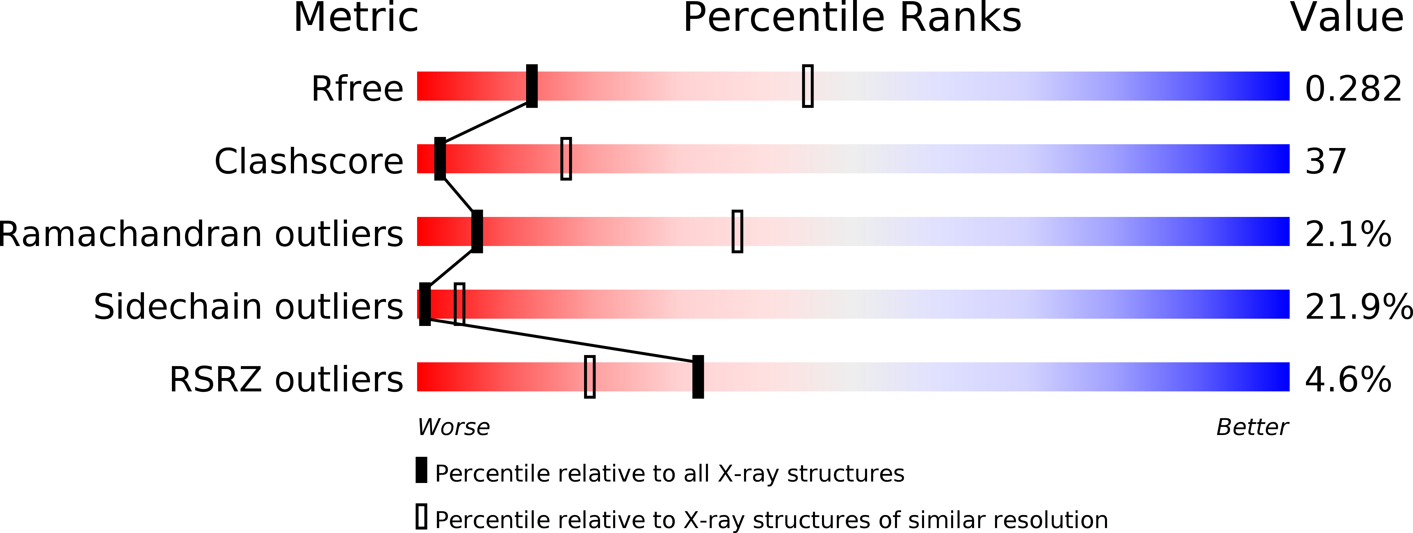
Resolution:
3.20 Å
R-Value Free:
0.27
R-Value Work:
0.24
R-Value Observed:
0.24
Space Group:
P 42 2 2
Macromolecular Entities
Structures with similar UniProt ID
Protein Blast
Polymer Type:polypeptide(L)
Molecule:DIPHTHERIA TOXIN REPRESSOR
Chain IDs:C (auth: A), D (auth: B), E (auth: C), F (auth: D)
Chain Length:225
Number of Molecules:4
Biological Source:Corynebacterium diphtheriae
Ligand Molecules
Primary Citation
Crystal structure of a cobalt-activated diphtheria toxin repressor-DNA complex reveals a metal-binding SH3-like domain.
J.Mol.Biol. 292 653 667 (1999)
PMID: 10497029 DOI: 10.1006/jmbi.1999.3073

Abstact

The diphtheria toxin repressor (DtxR) is the prototype of a family of iron-dependent regulator (IdeR) proteins, which are activated by divalent iron and bind DNA to prevent the transcription of downstream genes. In Corynebacterium diphtheriae, DtxR regulates not only the expression of diphtheria toxin encoded by a corynebacteriophage, but also of components of the siderophore-mediated iron-transport system. Here we report the crystal structure of wild-type DtxR, a 226 residue three-domain dimeric protein, activated by cobalt and bound to a 21 bp DNA duplex based on the consensus operator sequence. Two DtxR dimers surround the DNA duplex which is distorted compared to canonical B -DNA. The SH3-like third domain interacts with the metal at site 1 via the side-chains of Glu170 and Gln173, revealing for the first time a metal-binding function for this class of domains. The SH3-like domain is also in contact with the DNA-binding first domain and with the second, or dimerization, domain. The DNA-binding helices in the first domain are shifted by 3 to 5 A when compared to the apo-repressor, and fit into the major groove of the duplex bound. These shifts are due to a hinge-binding motion of the DNA-binding domain with respect to the dimerization domains of DtxR. The third domain might play a role in regulating this hinge motion.

Legend

Protein

Chemical

Disease

Primary Citation of related structures
Feedback Form
Name
Email
Institute
Feedback