Deposition Date
1998-10-27
Release Date
1999-04-27
Last Version Date
2024-05-22
Entry Detail
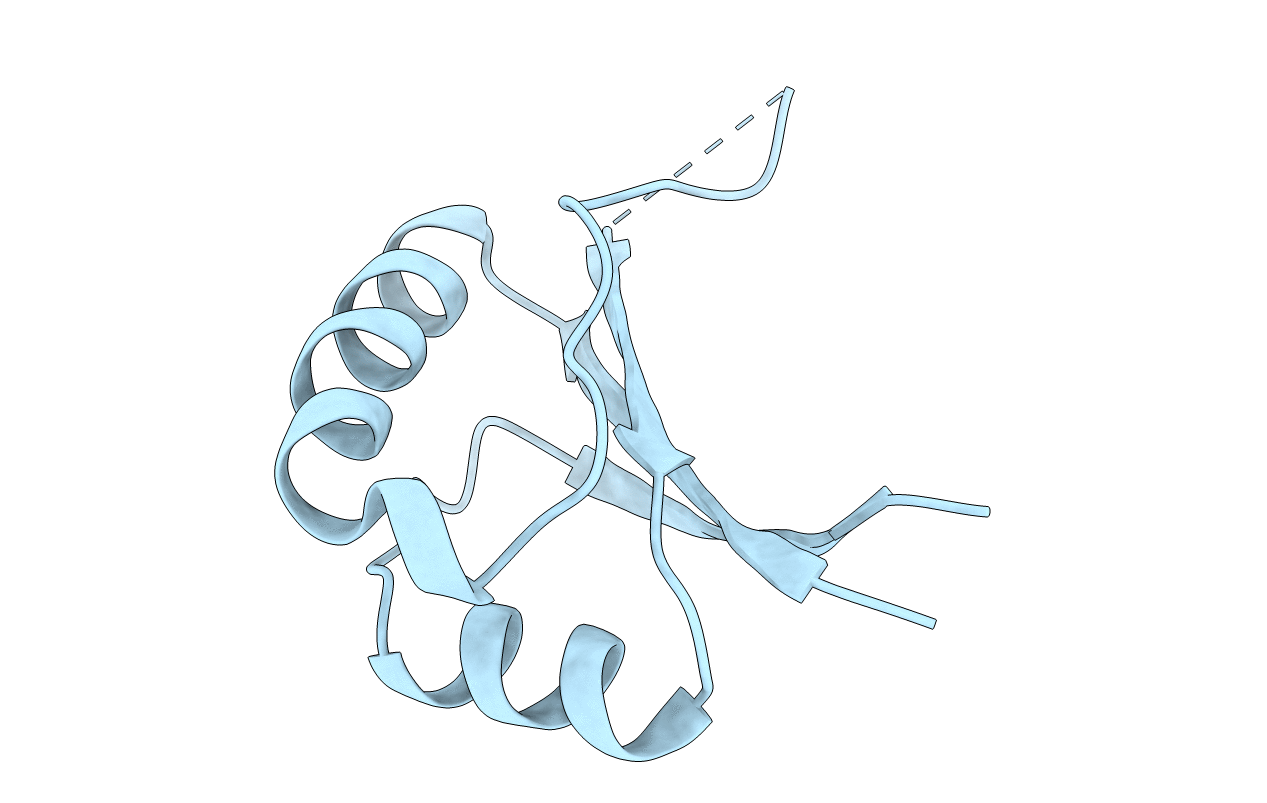
PDB ID:
1BY9
Keywords:
Title:
CRYSTAL STRUCTURE OF THE E2 DNA-BINDING DOMAIN FROM HUMAN PAPILLOMAVIRUS TYPE-16: IMPLICATIONS FOR ITS DNA BINDING-SITE SELECTION MECHANISM
Biological Source:
Source Organism(s):
Human papillomavirus type 16 (Taxon ID: 333760)
Expression System(s):
Method Details:
Experimental Method:
Resolution:
2.20 Å
R-Value Free:
0.22
R-Value Work:
0.19
R-Value Observed:
0.19
Space Group:
P 31 2 1