1a8y image
Deposition Date 1998-03-31
Release Date 1999-03-30
Last Version Date 2024-02-07
Entry Detail
PDB ID:
1A8Y
Title:
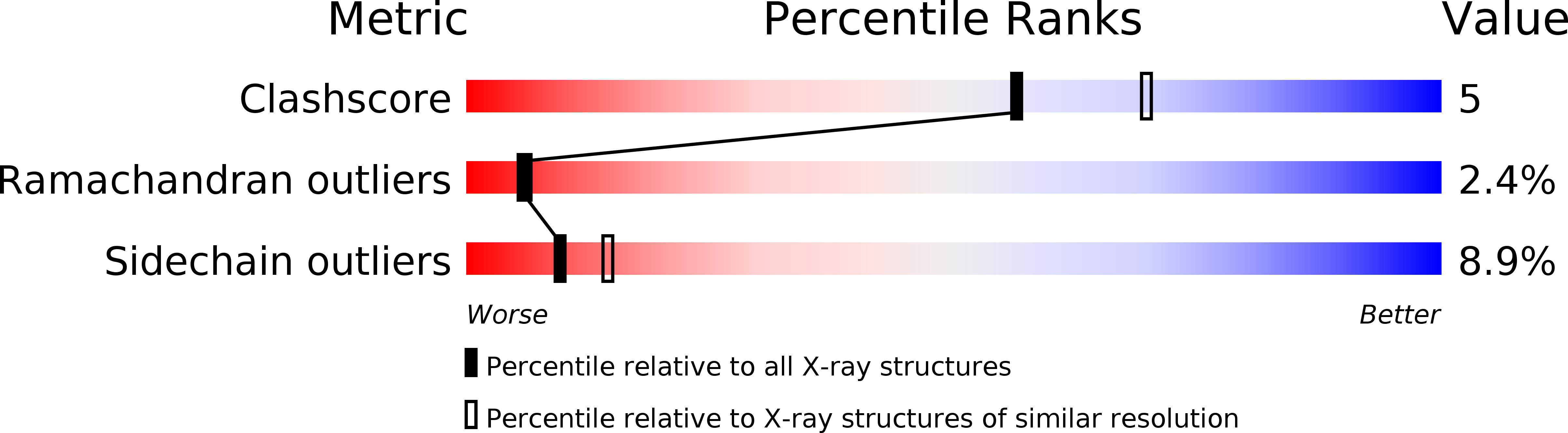
CRYSTAL STRUCTURE OF CALSEQUESTRIN FROM RABBIT SKELETAL MUSCLE SARCOPLASMIC RETICULUM AT 2.4 A RESOLUTION
Biological Source:
Source Organism:
Method Details:
Experimental Method:
Resolution:
2.40 Å
R-Value Free:
0.24
R-Value Work:
0.18
R-Value Observed:
0.18
Space Group:
C 2 2 21
Macromolecular Entities
Polymer Type:polypeptide(L)
Molecule:CALSEQUESTRIN
Gene (Uniprot):CASQ1
Chain IDs:A
Chain Length:367
Number of Molecules:1
Biological Source:Oryctolagus cuniculus
Primary Citation
Crystal structure of calsequestrin from rabbit skeletal muscle sarcoplasmic reticulum.
Nat.Struct.Biol. 5 476 483 (1998)
PMID: 9628486 DOI: 10.1038/nsb0698-476

Abstact

Calsequestrin, the major Ca2+ storage protein of muscle, coordinately binds and releases 40-50 Ca2+ ions per molecule for each contraction-relaxation cycle by an uncertain mechanism. We have determined the structure of rabbit skeletal muscle calsequestrin. Three very negative thioredoxin-like domains surround a hydrophilic center. Each monomer makes two extensive dimerization contacts, both of which involve the approach of many negative groups. This structure suggests a mechanism by which calsequestrin may achieve high capacity Ca2+ binding. The suggested mechanism involves Ca2+-induced collapse of the three domains and polymerization of calsequestrin monomers arising from three factors: N-terminal arm exchange, helix-helix contacts and Ca2+ cross bridges. This proposed structure-based mechanism accounts for the observed coupling of high capacity Ca2+ binding with protein precipitation.

Legend

Protein

Chemical

Disease

Primary Citation of related structures