Deposition Date
2005-04-05
Release Date
2005-06-14
Last Version Date
2024-11-06
Entry Detail
PDB ID:
1ZA3
Keywords:
Title:
The crystal structure of the YSd1 Fab bound to DR5
Biological Source:
Source Organism(s):
Homo sapiens (Taxon ID: 9606)
Expression System(s):
Method Details:
Experimental Method:
Resolution:
3.35 Å
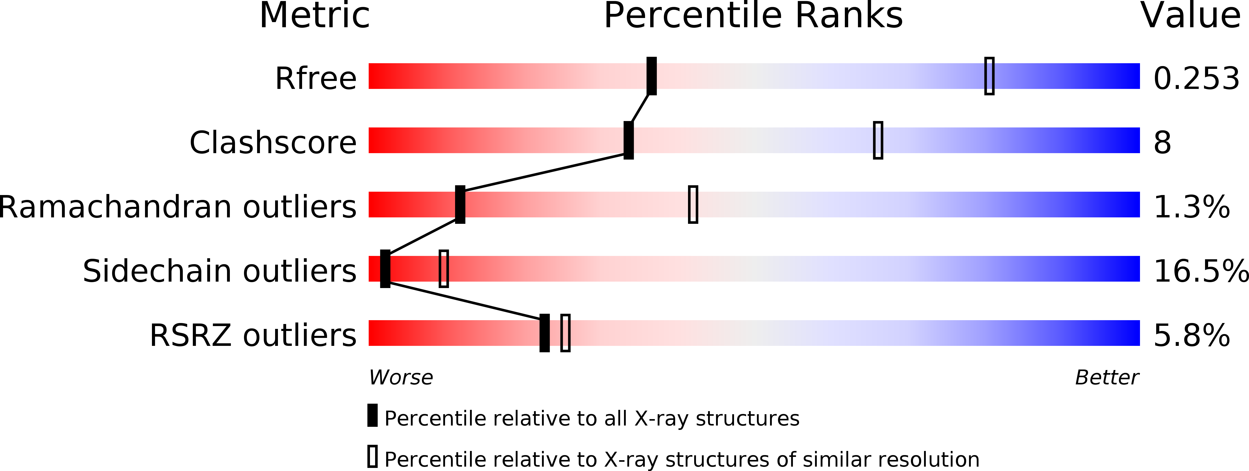
R-Value Free:
0.28
R-Value Work:
0.22
R-Value Observed:
0.22
Space Group:
P 32 2 1