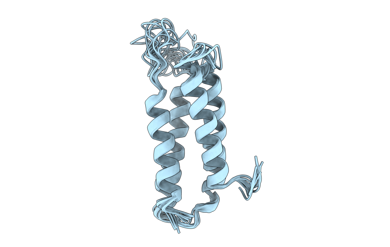
1ZZP image
Deposition Date 2005-06-14
Release Date 2005-08-30
Last Version Date 2024-05-22
Entry Detail
PDB ID:
1ZZP
Keywords:
Title:
Solution structure of the F-actin binding domain of Bcr-Abl/c-Abl
Biological Source:
Source Organism:
Homo sapiens (Taxon ID: 9606)
Method Details:
Experimental Method:
Conformers Calculated:
100
Conformers Submitted:
10
Selection Criteria:
structures with the lowest energy
Macromolecular Entities
Polymer Type:polypeptide(L)
Molecule:Proto-oncogene tyrosine-protein kinase ABL1
Gene (Uniprot):ABL1
Chain IDs:A
Chain Length:130
Number of Molecules:1
Biological Source:Homo sapiens
Ligand Molecules
Primary Citation
Structural Basis for the Cytoskeletal Association of Bcr-Abl/c-Abl.
Mol.Cell 19 461 473 (2005)
PMID: 16109371 DOI: 10.1016/j.molcel.2005.06.030

Abstact

The Bcr-Abl tyrosine kinase causes different forms of leukemia in humans. Depending on its position within the cell, Bcr-Abl differentially affects cellular growth. However, no structural and molecular details for the anticipated localization determinants are available. We present the NMR structure of the F-actin binding domain (FABD) of Bcr-Abl and its cellular counterpart c-Abl. The FABD forms a compact left-handed four-helix bundle in solution. We show that the nuclear export signal (NES) previously reported in this region is part of the hydrophobic core and nonfunctional in the intact protein. In contrast, we could identify the critical residues of helix alphaIII that are responsible for F-actin binding and cytoskeletal association. We propose that these interactions represent a major determinant for both Bcr-Abl and c-Abl localization.

Legend

Protein

Chemical

Disease

Primary Citation of related structures