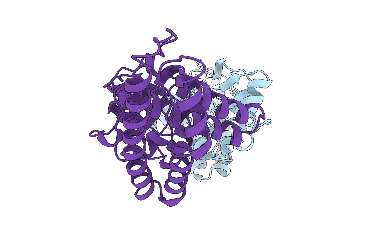
1ZYM image
Deposition Date 1996-05-21
Release Date 1996-12-07
Last Version Date 2024-02-14
Entry Detail
PDB ID:
1ZYM
Title:
AMINO TERMINAL DOMAIN OF ENZYME I FROM ESCHERICHIA COLI
Biological Source:
Source Organism:
Escherichia coli (Taxon ID: 562)
Host Organism:
Method Details:
Experimental Method:
Resolution:
2.50 Å
R-Value Free:
0.30
R-Value Work:
0.20
R-Value Observed:
0.20
Space Group:
P 21 21 21
Macromolecular Entities
Polymer Type:polypeptide(L)
Molecule:ENZYME I
Gene (Uniprot):ptsI
Chain IDs:A, B
Chain Length:258
Number of Molecules:2
Biological Source:Escherichia coli
Primary Citation
The first step in sugar transport: crystal structure of the amino terminal domain of enzyme I of the E. coli PEP: sugar phosphotransferase system and a model of the phosphotransfer complex with HPr.
Structure 4 861 872 (1996)
PMID: 8805571 DOI: 10.1016/S0969-2126(96)00092-5

Abstact

BACKGROUND The bacterial phosphoenolpyruvate (PEP): sugar phosphotransferase system (PTS) transports exogenous hexose sugars through the membrane and tightly couples transport with phosphoryl transfer from PEP to the sugar via several phosphoprotein intermediates. The phosphate group is first transferred to enzyme I, second to the histidine-containing phosphocarrier protein HPr, and then to one of a number of sugar-specific enzymes II. The structures of several HPrs and enzymes IIA are known. Here we report the structure of the N-terminal half of enzyme I from Escherichia coli (EIN). RESULTS The crystal structure of EIN (MW approximately 30 kDa) has been determined and refined at 2.5 A resolution. It has two distinct structural subdomains; one contains four alpha helices arranged as two hairpins in a claw-like conformation. The other consists of a beta sandwich containing a three-stranded antiparallel beta sheet and a four-stranded parallel beta sheet, together with three short alpha helices. Plausible models of complexes between EIN and HPr can be made without assuming major structural changes in either protein. CONCLUSIONS The alpha/beta subdomain of EIN is topologically similar to the phosphohistidine domain of the enzyme pyruvate phosphate dikinase, which is phosphorylated by PEP on a histidyl residue but does not interact with HPr. It is therefore likely that features of this subdomain are important in the autophosphorylation of enzyme I. The helical subdomain of EIN is not found in pyruvate phosphate dikinase; this subdomain is therefore more likely to be involved in phosphoryl transfer to HPr.

Legend

Protein

Chemical

Disease

Primary Citation of related structures