1ZYE image
Deposition Date 2005-06-10
Release Date 2005-09-20
Last Version Date 2023-10-25
Entry Detail
PDB ID:
1ZYE
Keywords:
Title:
Crystal structure analysis of Bovine Mitochondrial Peroxiredoxin III
Biological Source:
Source Organism:
Bos taurus (Taxon ID: 9913)
Host Organism:
Method Details:
Experimental Method:
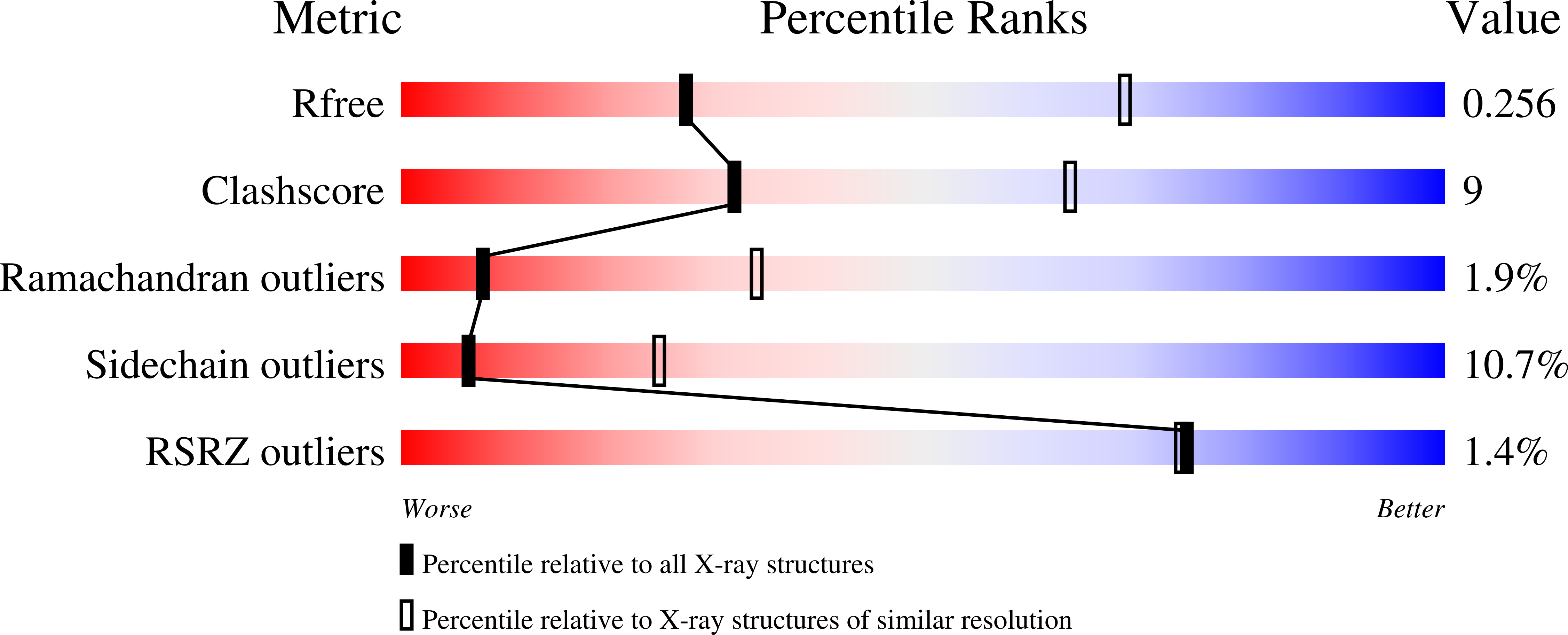
Resolution:
3.30 Å
R-Value Free:
0.26
R-Value Work:
0.22
R-Value Observed:
0.22
Space Group:
C 1 2 1
Macromolecular Entities
Structures with similar UniProt ID
Protein Blast
Polymer Type:polypeptide(L)
Molecule:Thioredoxin-dependent peroxide reductase
Gene (Uniprot):PRDX3
Mutagens:C168S
Chain IDs:A, B, C, D, E, F, G, H, I, J, K, L
Chain Length:220
Number of Molecules:12
Biological Source:Bos taurus
Primary Citation
Bovine Mitochondrial Peroxiredoxin III Forms a Two-Ring Catenane
Structure 13 1661 1664 (2005)
PMID: 16271889 DOI: 10.1016/j.str.2005.07.021

Abstact

A crystal structure is reported for the C168S mutant of a typical 2-Cys peroxiredoxin III (Prx III) from bovine mitochondria at a resolution of 3.3 A. Prx III is present as a two-ring catenane comprising two interlocking dodecameric toroids that are assembled from basic dimeric units. Each ring has an external diameter of 150 A and encompasses a central cavity that is 70 A in width. The concatenated dodecamers are inclined at an angle of 55 degrees, which provides a large contact surface between the rings. Dimer-dimer contacts involved in toroid formation are hydrophobic in nature, whereas the 12 areas of contact between interlocked rings arise from polar interactions. These two major modes of subunit interaction provide important insights into possible mechanisms of catenane formation.

Legend

Protein

Chemical

Disease

Primary Citation of related structures