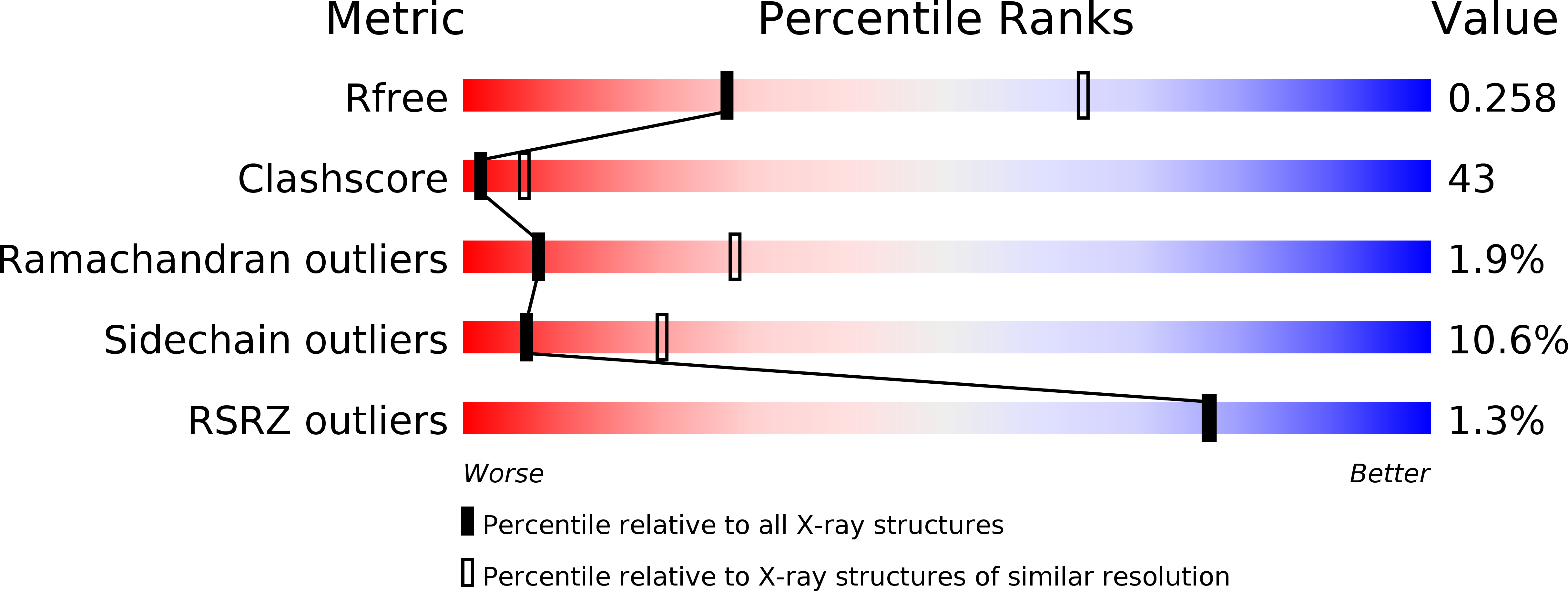
1ZY0 image
Deposition Date 2005-06-09
Release Date 2005-09-27
Last Version Date 2023-08-23
Entry Detail
PDB ID:
1ZY0
Keywords:
Title:
X-ray structure of peptide deformylase from Arabidopsis thaliana (AtPDF1A); crystals grown in PEG-6000
Biological Source:
Source Organism:
Method Details:
Experimental Method:
Resolution:
2.90 Å
R-Value Free:
0.28
R-Value Work:
0.24
Space Group:
P 21 21 21
Macromolecular Entities
Polymer Type:polypeptide(L)
Molecule:Peptide deformylase, mitochondrial
Gene (Uniprot):PDF1A
Chain IDs:A, B
Chain Length:197
Number of Molecules:2
Biological Source:Arabidopsis thaliana
Ligand Molecules
Primary Citation
The crystal structure of mitochondrial (Type 1A) peptide deformylase provides clear guidelines for the design of inhibitors specific for the bacterial forms
J.Biol.Chem. 280 42315 42324 (2005)
PMID: 16192279 DOI: 10.1074/jbc.M507155200

Abstact

Peptide deformylase (PDF) inhibitors have a strong potential to be used as a new class of antibiotics. However, recent studies have shown that the mitochondria of most eukaryotes, including humans, contain an essential PDF, PDF1A. The crystal structure of the Arabidopsis thaliana PDF1A (AtPDF1A), considered representative of PDF1As in general, has been determined. This structure displays several similarities to that of known bacterial PDFs. AtPDF1A behaves as a dimer, with the C-terminal residues responsible for linking the two subunits. This arrangement is similar to that of Leptospira interrogans PDF, the only other dimeric PDF identified to date. AtPDF1A is the first PDF for which zinc has been identified as the catalytic ion. However, the zinc binding pocket does not differ from the binding pockets of PDFs with iron rather than zinc. The crystal structure of AtPDF1A in complex with a substrate analog revealed that the substrate binding pocket of PDF1A displays strong modifications. The S1' binding pocket is significantly narrower, due to the creation of a floor from residues present in all PDF1As but not in bacterial PDFs. A true S3' pocket is created by the residues of a helical CD-loop, which is very long in PDF1As. Finally, these modified substrate binding pockets modify the position of the substrate in the active site. These differences provide guidelines for the design of bacterial PDF inhibitors that will not target mitochondrial PDFs.

Legend

Protein

Chemical

Disease

Primary Citation of related structures