1ZWJ image
Deposition Date 2005-06-03
Release Date 2005-06-14
Last Version Date 2023-08-23
Entry Detail
PDB ID:
1ZWJ
Title:
X-ray structure of galt-like protein from arabidopsis thaliana AT5G18200
Biological Source:
Source Organism:
Host Organism:
Method Details:
Experimental Method:
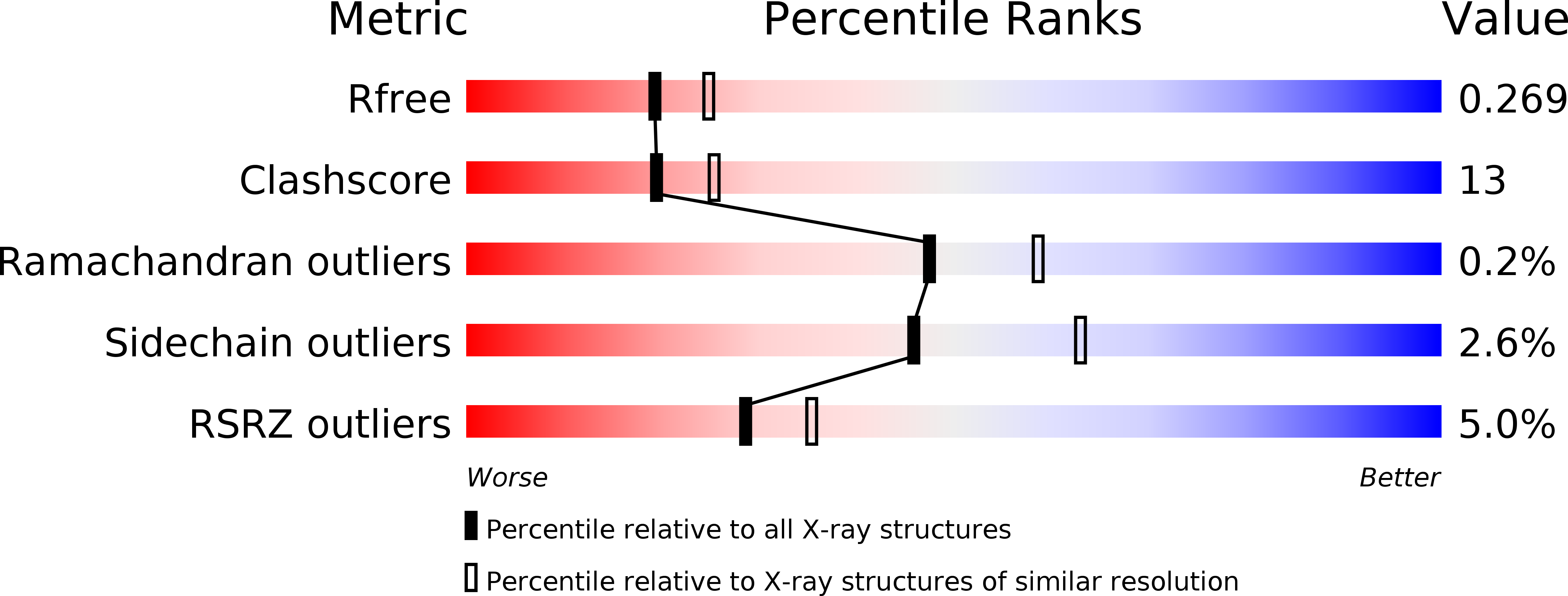
Resolution:
2.30 Å
R-Value Free:
0.26
R-Value Work:
0.21
Space Group:
P 21 21 21
Macromolecular Entities
Polymer Type:polypeptide(L)
Molecule:putative galactose-1-phosphate uridyl transferase
Gene (Uniprot):MRG7.16
Chain IDs:A, B
Chain Length:351
Number of Molecules:2
Biological Source:Arabidopsis thaliana
Ligand Molecules
Primary Citation
Structure and Mechanism of an ADP-Glucose Phosphorylase from Arabidopsis thaliana
Biochemistry 45 3154 3162 (2006)
PMID: 16519510 DOI: 10.1021/bi052232m

Abstact

The X-ray crystal structure of the At5g18200.1 protein has been determined to a nominal resolution of 2.30 A. The structure has a histidine triad (HIT)-like fold containing two distinct HIT-like motifs. The sequence of At5g18200.1 indicates a distant family relationship to the Escherichia coli galactose-1-P uridylyltransferase (GalT): the determined structure of the At5g18200.1 protein confirms this relationship. The At5g18200.1 protein does not demonstrate GalT activity but instead catalyzes adenylyl transfer in the reaction of ADP-glucose with various phosphates. The best acceptor among those evaluated is phosphate itself; thus, the At5g18200.1 enzyme appears to be an ADP-glucose phosphorylase. The enzyme catalyzes the exchange of (14)C between ADP-[(14)C]glucose and glucose-1-P in the absence of phosphate. The steady state kinetics of exchange follows the ping-pong bi-bi kinetic mechanism, with a k(cat) of 4.1 s(-)(1) and K(m) values of 1.4 and 83 microM for ADP-[(14)C]glucose and glucose-1-P, respectively, at pH 8.5 and 25 degrees C. The overall reaction of ADP-glucose with phosphate to produce ADP and glucose-1-P follows ping-pong bi-bi steady state kinetics, with a k(cat) of 2.7 s(-)(1) and K(m) values of 6.9 and 90 microM for ADP-glucose and phosphate, respectively, at pH 8.5 and 25 degrees C. The kinetics are consistent with a double-displacement mechanism that involves a covalent adenylyl-enzyme intermediate. The X-ray crystal structure of this intermediate was determined to 1.83 A resolution and shows the AMP group bonded to His(186). The value of K(eq) in the direction of ADP and glucose-1-P formation is 5.0 at pH 7.0 and 25 degrees C in the absence of a divalent metal ion, and it is 40 in the presence of 1 mM MgCl(2).

Legend

Protein

Chemical

Disease

Primary Citation of related structures