1ZW2 image
Deposition Date 2005-06-03
Release Date 2005-09-20
Last Version Date 2023-08-23
Entry Detail
PDB ID:
1ZW2
Keywords:
Title:
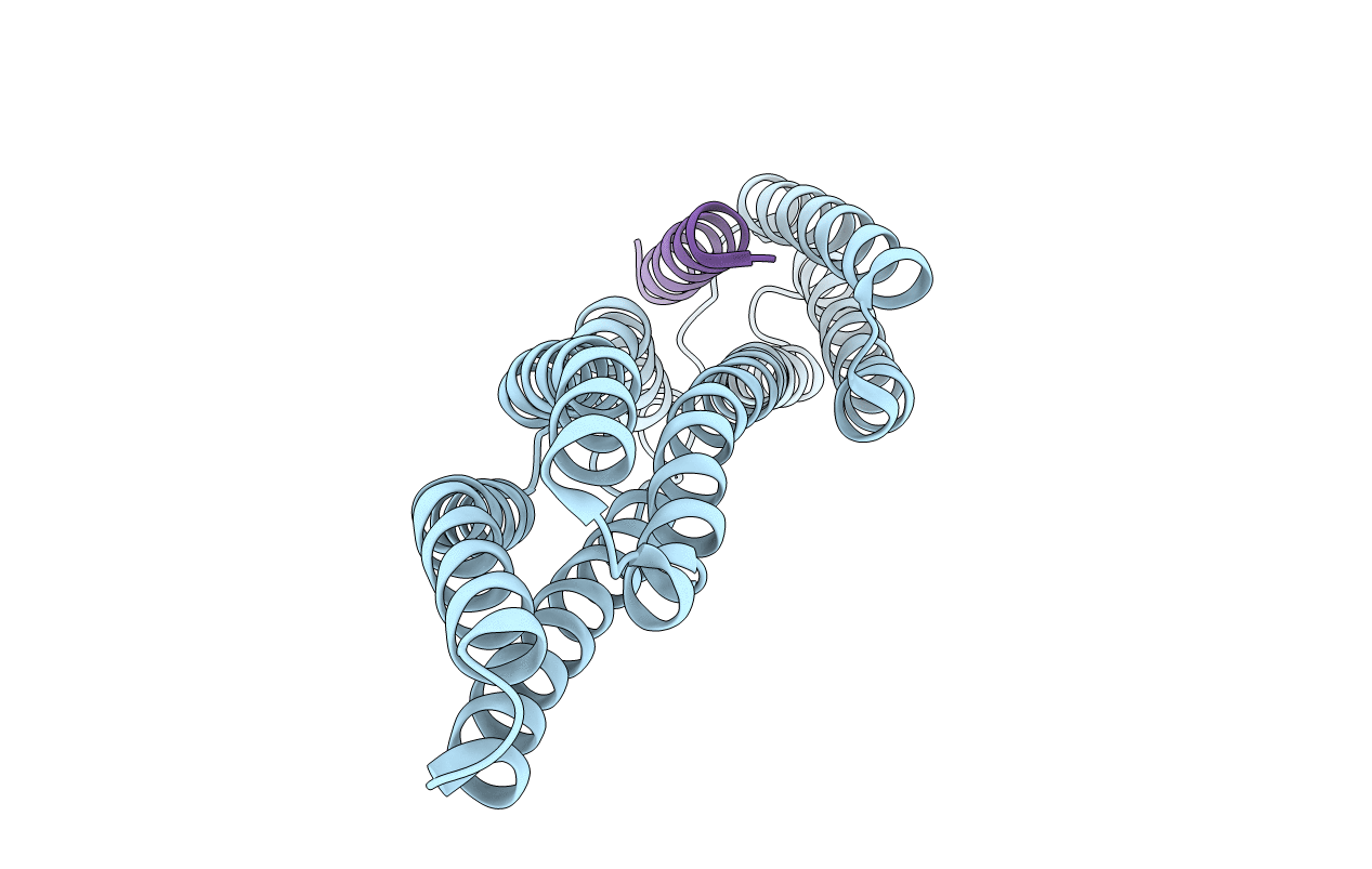
Vinculin Head (0-258) in Complex with the Talin Rod residues 2345-2369
Biological Source:
Source Organism:
Gallus gallus (Taxon ID: 9031)
(Taxon ID: )
Method Details:
Experimental Method:
Resolution:
2.10 Å
R-Value Free:
0.29
R-Value Work:
0.25
R-Value Observed:
0.25
Space Group:
P 21 21 2
Macromolecular Entities
Polymer Type:polypeptide(L)
Molecule:Vinculin
Gene (Uniprot):VCL
Chain IDs:A
Chain Length:279
Number of Molecules:1
Biological Source:Gallus gallus
Polymer Type:polypeptide(L)
Molecule:talin
Chain IDs:B
Chain Length:25
Number of Molecules:1
Biological Source:
Primary Citation
Mapping and consensus sequence identification for multiple vinculin binding sites within the talin rod
J.Biol.Chem. 280 37217 37224 (2005)
PMID: 16135522 DOI: 10.1074/jbc.M508060200

Abstact

The interaction between the cytoskeletal proteins talin and vinculin plays a key role in integrin-mediated cell adhesion and migration. Three vinculin binding sites (VBS1-3) have previously been identified in the talin rod using a yeast two-hybrid assay. To extend these studies, we spot-synthesized a series of peptides spanning all the alpha-helical regions predicted for the talin rod and identified eight additional VBSs, two of which overlap key functional regions of the rod, including the integrin binding site and C-terminal actin binding site. The talin VBS alpha-helices bind to a hydrophobic cleft in the N-terminal vinculin Vd1 domain. We have defined the specificity of this interaction by spot-synthesizing a series of 25-mer talin VBS1 peptides containing substitutions with all the commonly occurring amino acids. The consensus for recognition is LXXAAXXVAXX- VXXLIXXA with distinct classes of hydrophobic side chains at positions 1, 4, 5, 8, 9, 12, 15, and 16 required for vinculin binding. Positions 1, 8, 12, 15, and 16 require an aliphatic residue and will not tolerate alanine, whereas positions 4, 5, and 9 are less restrictive. These preferences are common to all 11 VBS sequences with a minor variation occurring in one case. A crystal structure of this variant VBS peptide in complex with the vinculin Vd1 domain reveals a subtly different mode of vinculin binding.

Legend

Protein

Chemical

Disease

Primary Citation of related structures