1ZVW image
Entry Detail
PDB ID:
1ZVW
Keywords:
Title:
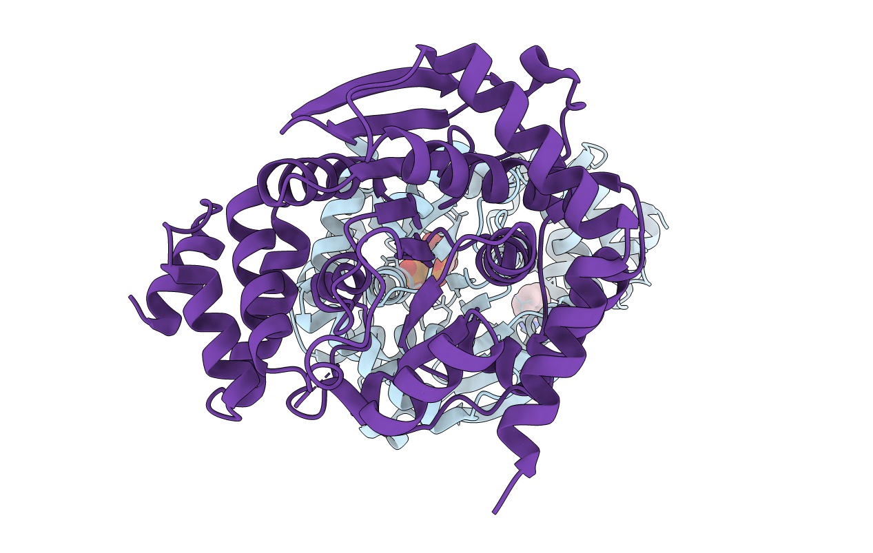
The Crystal Structure of TrpD (Rv2192c) from Mycobacterium tuberculosis in Complex with PRPP and Magnesium
Biological Source:
Host Organism:
PDB Version:
Deposition Date:
2005-06-02
Release Date:
2006-01-17
Method Details:
Experimental Method:
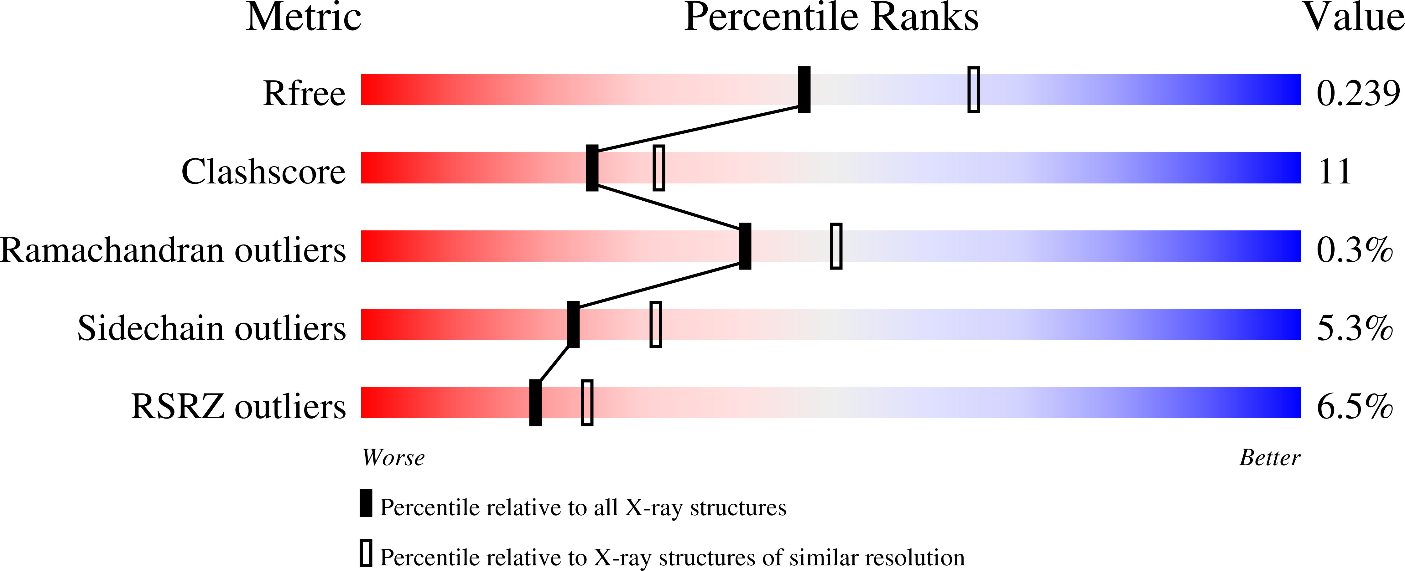
Resolution:
2.30 Å
R-Value Free:
0.25
R-Value Work:
0.19
R-Value Observed:
0.19
Space Group:
P 21 21 21
Macromolecular Entities
Polymer Type:polypeptide(L)
Description:Anthranilate phosphoribosyltransferase
Chain IDs:A, B
Chain Length:378
Number of Molecules:2
Biological Source:Mycobacterium tuberculosis
Modified Residue
Compound ID Chain ID Parent Comp ID Details 2D Image
MSE A MET SELENOMETHIONINE
Primary Citation
The Crystal Structure of TrpD, a Metabolic Enzyme Essential for Lung Colonization by Mycobacterium tuberculosis, in Complex with its Substrate Phosphoribosylpyrophosphate.
J.Mol.Biol. 355 784 797 (2006)
PMID: 16337227 DOI: 10.1016/j.jmb.2005.11.016

Abstact

Mycobacterium tuberculosis, the cause of tuberculosis, presents a major threat to human health worldwide. Biosynthetic enzymes that are essential for the survival of the bacterium, especially in activated macrophages, are important potential drug targets. Although the tryptophan biosynthesis pathway is thought to be non-essential for many pathogens, this appears not to be the case for M.tuberculosis, where a trpD gene knockout fails to cause disease in mice. We therefore chose the product of the trpD gene, anthranilate phosphoribosyltransferase, which catalyses the second step in tryptophan biosynthesis, for structural analysis. The structure of TrpD from M.tuberculosis was solved by X-ray crystallography, at 1.9 A resolution for the native enzyme (R = 0.191, Rfree = 0.230) and at 2.3 A resolution for the complex with its substrate phosphoribosylpyrophosphate (PRPP) and Mg2+ (R = 0.194, Rfree = 0.255). The enzyme is folded into two domains, separated by a hinge region. PRPP binds in the C-terminal domain, together with a pair of Mg ions. In the substrate complex, two flexible loops change conformation compared with the apo protein, to close over the PRPP and to complete an extensive network of hydrogen-bonded interactions. A nearby pocket, adjacent to the hinge region, is postulated by in silico docking as the binding site for anthranilate. A bound molecule of benzamidine, which was essential for crystallization and is also found in the hinge region, appears to reduce flexibility between the two domains.

Legend

Protein

Chemical

Disease

Primary Citation of related structures