1ZUN image
Deposition Date 2005-05-31
Release Date 2006-01-17
Last Version Date 2024-11-13
Entry Detail
PDB ID:
1ZUN
Keywords:
Title:
Crystal Structure of a GTP-Regulated ATP Sulfurylase Heterodimer from Pseudomonas syringae
Biological Source:
Expression System(s):
Method Details:
Experimental Method:
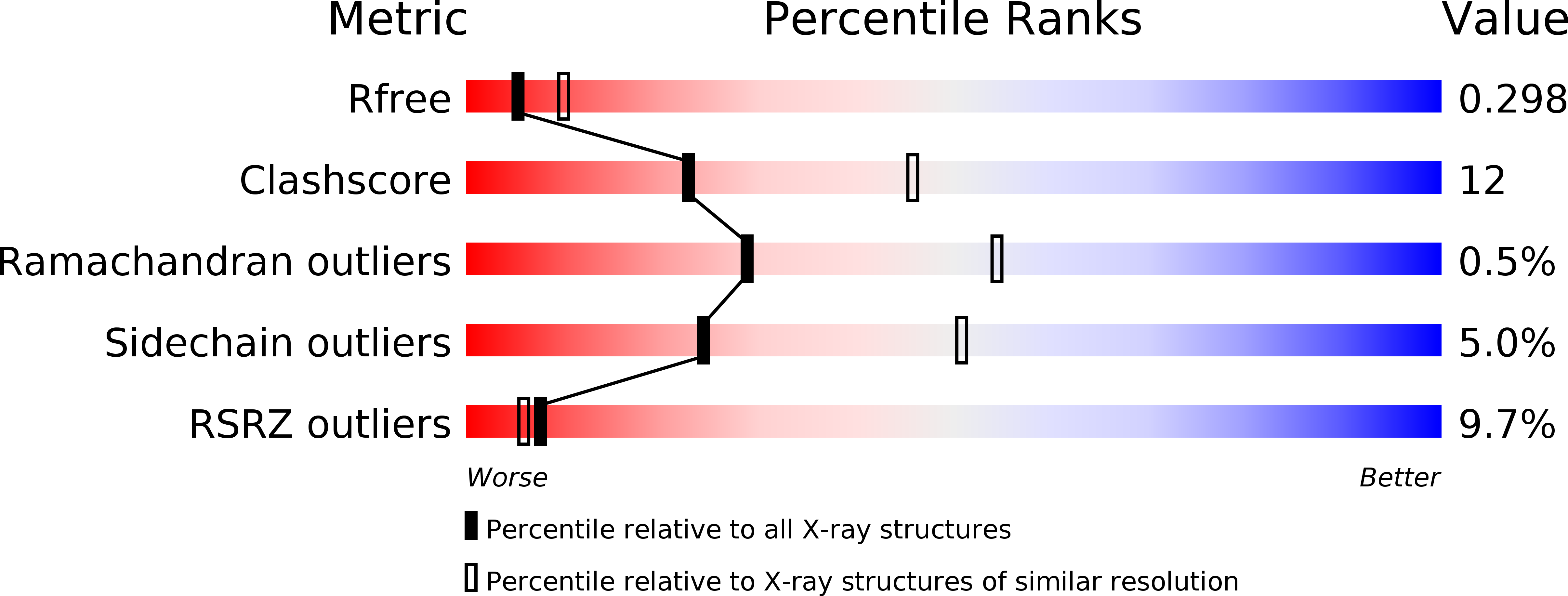
Resolution:
2.70 Å
R-Value Free:
0.27
R-Value Work:
0.22
R-Value Observed:
0.22
Space Group:
H 3
Macromolecular Entities
Structures with similar UniProt ID
Protein Blast
Polymer Type:polypeptide(L)
Molecule:Sulfate adenylyltransferase subunit 2
Gene (Uniprot):cysD
Chain IDs:A
Chain Length:325
Number of Molecules:1
Biological Source:Pseudomonas syringae
Structures with similar UniProt ID
Protein Blast
Polymer Type:polypeptide(L)
Molecule:sulfate adenylate transferase, subunit 1/adenylylsulfate kinase
Chain IDs:B
Chain Length:434
Number of Molecules:1
Biological Source:Pseudomonas syringae pv. tomato str. DC3000
Modified Residue
Compound ID Chain ID Parent Comp ID Details 2D Image
MSE A MET SELENOMETHIONINE
Primary Citation
Molecular basis for g protein control of the prokaryotic ATP sulfurylase.
Mol.Cell 21 109 122 (2006)
PMID: 16387658 DOI: 10.1016/j.molcel.2005.10.034

Abstact

Sulfate assimilation is a critical component of both primary and secondary metabolism. An essential step in this pathway is the activation of sulfate through adenylation by the enzyme ATP sulfurylase (ATPS), forming adenosine 5'-phosphosulfate (APS). Proteobacterial ATPS overcomes this energetically unfavorable reaction by associating with a regulatory G protein, coupling the energy of GTP hydrolysis to APS formation. To discover the molecular basis of this unusual role for a G protein, we biochemically characterized and solved the X-ray crystal structure of a complex between Pseudomonas syringae ATPS (CysD) and its associated regulatory G protein (CysN). The structure of CysN*D shows the two proteins in tight association; however, the nucleotides bound to each subunit are spatially segregated. We provide evidence that conserved switch motifs in the G domain of CysN allosterically mediate interactions between the nucleotide binding sites. This structure suggests a molecular mechanism by which conserved G domain architecture is used to energetically link GTP turnover to the production of an essential metabolite.

Legend

Protein

Chemical

Disease

Primary Citation of related structures
Feedback Form
Name
Email
Institute
Feedback