
Deposition Date
1996-11-15
Release Date
1997-06-05
Last Version Date
2022-03-02
Entry Detail
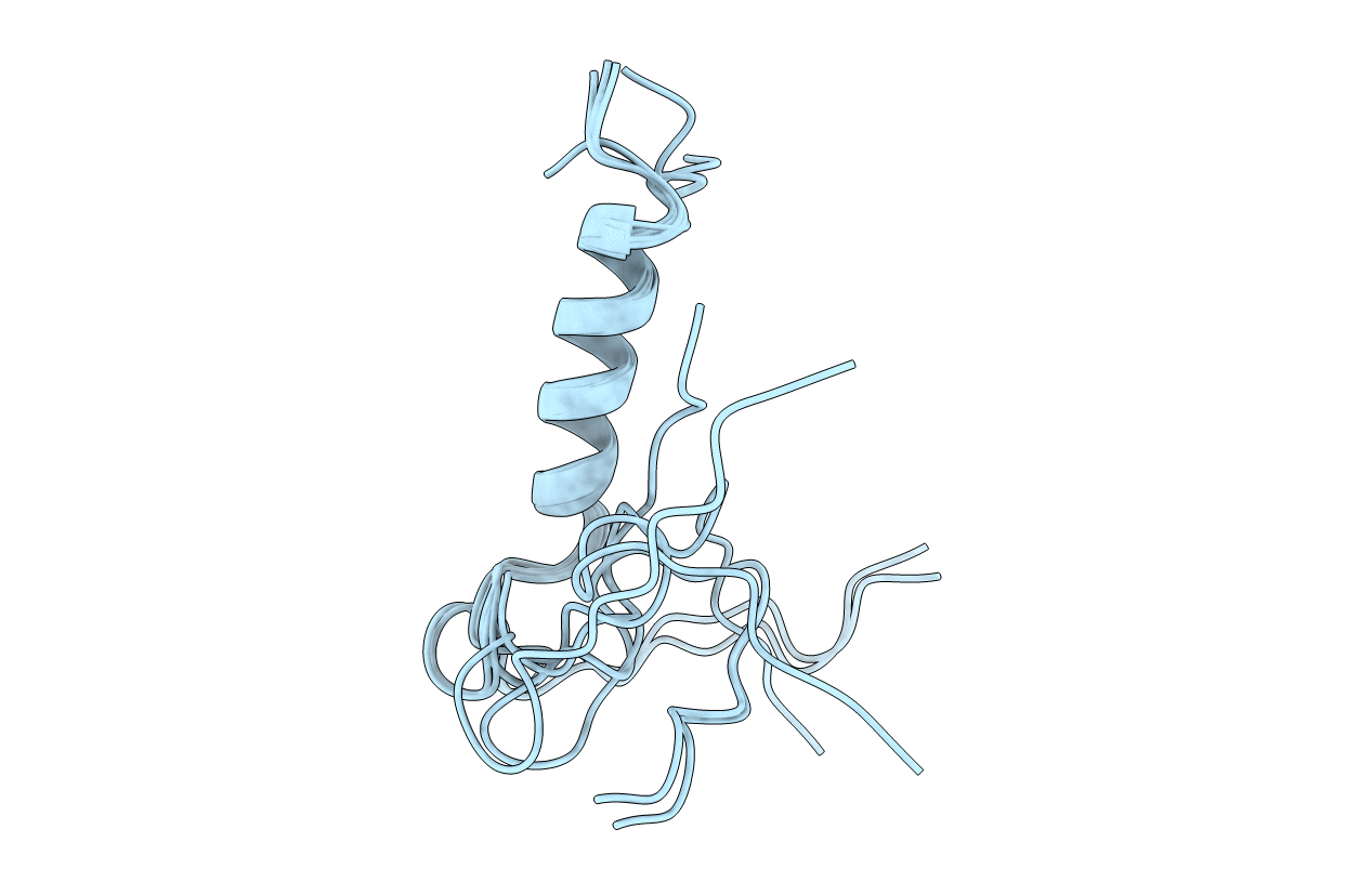
PDB ID:
1ZTO
Keywords:
Title:
INACTIVATION GATE OF POTASSIUM CHANNEL RCK4, NMR, 8 STRUCTURES
Biological Source:
Source Organism(s):
Homo sapiens (Taxon ID: 9606)
Method Details:
