1ZT7 image
Deposition Date 2005-05-26
Release Date 2005-10-18
Last Version Date 2024-11-06
Entry Detail
PDB ID:
1ZT7
Keywords:
Title:
crystal structure of class I MHC H-2Kk in complex with a nonapeptide
Biological Source:
Source Organism:
Mus musculus (Taxon ID: 10090)
Host Organism:
Method Details:
Experimental Method:
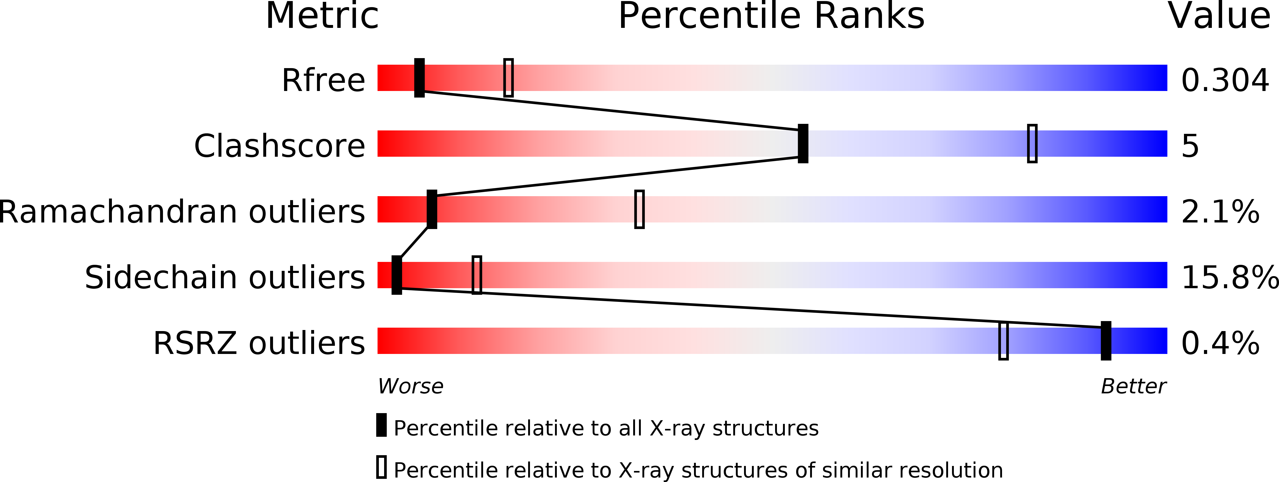
Resolution:
3.00 Å
R-Value Free:
0.31
R-Value Work:
0.23
R-Value Observed:
0.23
Space Group:
P 1 2 1
Macromolecular Entities
Structures with similar UniProt ID
Protein Blast
Polymer Type:polypeptide(L)
Molecule:H-2 class I histocompatibility antigen, K-K alpha chain
Gene (Uniprot):H2-K1
Chain IDs:A, C
Chain Length:276
Number of Molecules:2
Biological Source:Mus musculus
Structures with similar UniProt ID
Protein Blast
Polymer Type:polypeptide(L)
Molecule:Beta-2-microglobulin
Gene (Uniprot):B2m
Chain IDs:B, D
Chain Length:100
Number of Molecules:2
Biological Source:Mus musculus
Polymer Type:polypeptide(L)
Molecule:SV40 epitope, SEFLLEKRI
Chain IDs:E (auth: P), F (auth: Q)
Chain Length:9
Number of Molecules:2
Biological Source:
Primary Citation
The H-2Kk MHC peptide-binding groove anchors the backbone of an octameric antigenic peptide in an unprecedented mode.
J Immunol. 175 3819 3825 (2005)
PMID: 16148128 DOI: 10.4049/jimmunol.175.6.3819

Abstact

A wealth of data has accumulated on the structure of mouse MHC class I (MHCI) molecules encoded by the H-2(b) and H-2(d) haplotypes. In contrast, there is a dearth of structural data regarding H-2(k)-encoded molecules. Therefore, the structures of H-2K(k) complexed to an octameric peptide from influenza A virus (HA(259-266)) and to a nonameric peptide from SV40 (SV40(560-568)) have been determined by x-ray crystallography at 2.5 and 3.0 A resolutions, respectively. The structure of the H-2K(k)-HA(259-266) complex reveals that residues located on the floor of the peptide-binding groove contact directly the backbone of the octameric peptide and force it to lie deep within the H-2K(k) groove. This unprecedented mode of peptide binding occurs despite the presence of bulky residues in the middle of the floor of the H-2K(k) peptide-binding groove. As a result, the Calpha atoms of peptide residues P5 and P6 are more buried than the corresponding residues of H-2K(b)-bound octapeptides, making them even less accessible to TCR contact. When bound to H-2K(k), the backbone of the SV40(560-568) nonapeptide bulges out of the peptide-binding groove and adopts a conformation reminiscent of that observed for peptides bound to H-2L(d). This structural convergence occurs despite the totally different architectures of the H-2L(d) and H-2K(k) peptide-binding grooves. Therefore, these two H-2K(k)-peptide complexes provide insights into the mechanisms through which MHC polymorphism outside primary peptide pockets influences the conformation of the bound peptides and have implications for TCR recognition and vaccine design.

Legend

Protein

Chemical

Disease

Primary Citation of related structures