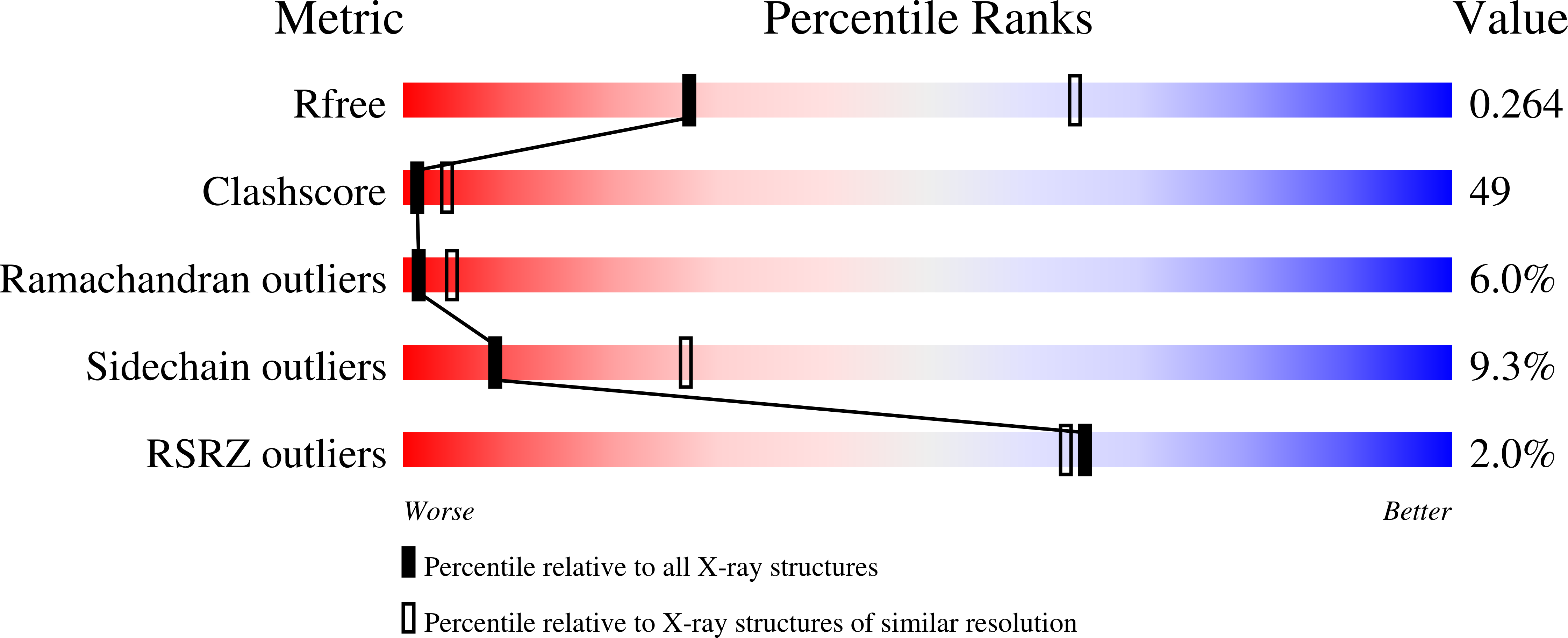
1ZSH image
Deposition Date 2005-05-24
Release Date 2006-01-31
Last Version Date 2023-08-23
Entry Detail
PDB ID:
1ZSH
Title:
Crystal structure of bovine arrestin-2 in complex with inositol hexakisphosphate (IP6)
Biological Source:
Source Organism:
Bos taurus (Taxon ID: 9913)
Host Organism:
Method Details:
Experimental Method:
Resolution:
2.90 Å
R-Value Free:
0.26
R-Value Work:
0.25
Space Group:
P 32 2 1
Macromolecular Entities
Polymer Type:polypeptide(L)
Molecule:Beta-arrestin 1
Gene (Uniprot):ARRB1
Chain IDs:A
Chain Length:418
Number of Molecules:1
Biological Source:Bos taurus
Primary Citation
Nonvisual arrestin oligomerization and cellular localization are regulated by inositol hexakisphosphate binding
J.Biol.Chem. 281 9812 9823 (2006)
PMID: 16439357 DOI: 10.1074/jbc.M512703200

Abstact

Interactions between arrestins and phosphoinositides have been reported to regulate multiple membrane-associated signaling and trafficking events including clathrin-mediated endocytosis and light adaptation in Drosophila. Arrestins have been proposed to have nuclear and cytosolic functions as well, although the ligand dependence of these functions has not been investigated. Here we characterize the structural, molecular, and cellular interactions between arrestin-2 and inositol hexakisphosphate (inositol 1,2,3,4,5,6-hexakisphosphate (IP(6))). The crystal structure of the arrestin-2.IP(6) complex was solved to 2.9 A with crystal lattice contacts suggesting two sites on a protein monomer mediating IP(6) binding. Mutagenesis coupled to isothermal titration calorimetry and tritiated IP(6) binding assays confirmed two-site binding with a low affinity IP(6)-binding site in the N-domain and a high affinity site in the C-domain. Native gel electrophoresis, gel filtration, and analytical ultracentrifugation demonstrated the ability of IP(6) to promote arrestin-2 oligomerization via the two crystallographically defined ligand-binding locations. In addition, analysis in mammalian cells revealed that arrestin-2 not only undergoes homo-oligomerization, but it can also hetero-oligomerize with arrestin-3 in a manner that depends on IP(6)-binding sites. Mutation of either IP(6)-binding site in arrestin-2 disrupted oligomerization while interactions with known binding partners including clathrin, AP-2, and ERK2 were maintained. Subcellular localization studies showed that arrestin-2 oligomers are primarily cytoplasmic, whereas arrestin-2 monomers displayed increased nuclear localization. Thus, by promoting cytosolic oligomerization, IP(6) binding is proposed to be a negative regulator of interactions of arrestin with plasma membrane and nuclear signaling proteins.

Legend

Protein

Chemical

Disease

Primary Citation of related structures