1ZS4 image
Deposition Date 2005-05-23
Release Date 2005-08-23
Last Version Date 2024-02-14
Entry Detail
PDB ID:
1ZS4
Title:
Structure of bacteriophage lambda cII protein in complex with DNA
Biological Source:
Source Organism:
Method Details:
Experimental Method:
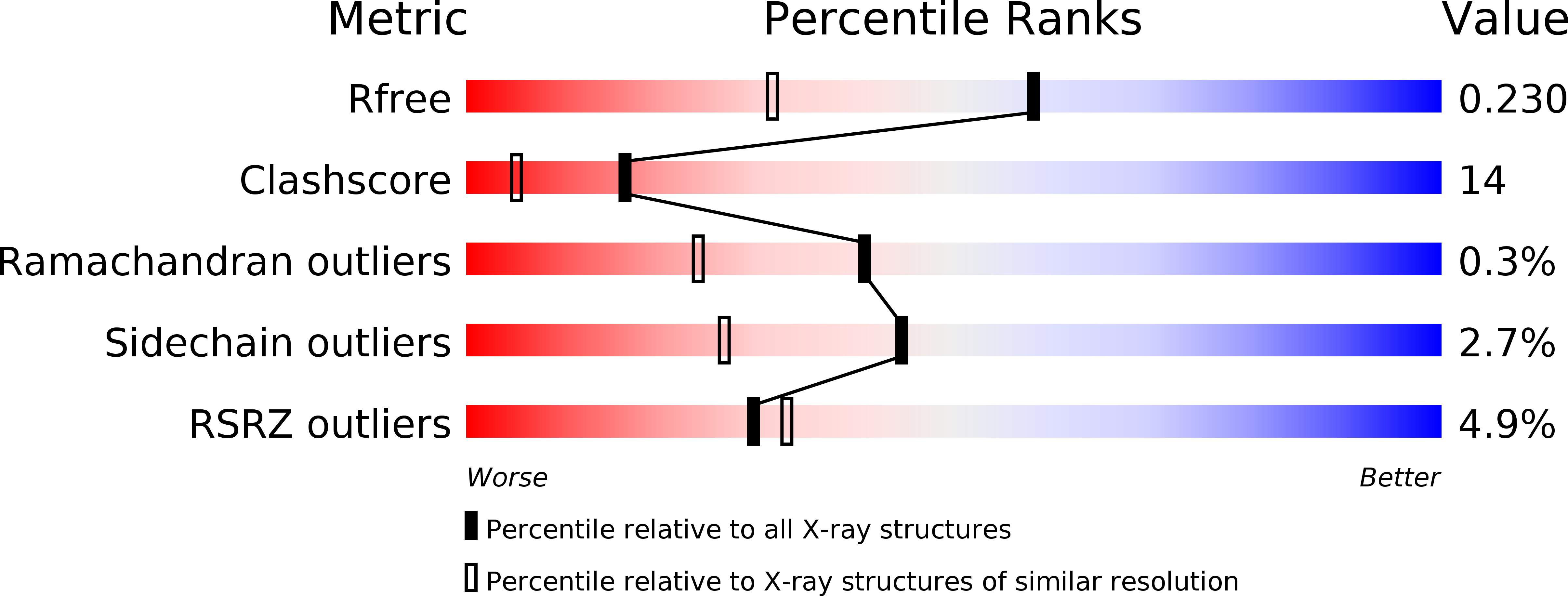
Resolution:
1.70 Å
R-Value Free:
0.22
R-Value Work:
0.20
Space Group:
P 1 21 1
Macromolecular Entities
Structures with similar UniProt ID
Protein Blast
Polymer Type:polypeptide(L)
Molecule:Regulatory protein CII
Gene (Uniprot):cII
Chain IDs:C (auth: A), D (auth: B), E (auth: C), F (auth: D)
Chain Length:83
Number of Molecules:4
Biological Source:Enterobacteria phage lambda
Polymer Type:polydeoxyribonucleotide
Molecule:DNA - 27mer
Chain IDs:B (auth: T)
Chain Length:27
Number of Molecules:1
Biological Source:
Polymer Type:polydeoxyribonucleotide
Molecule:DNA - 27mer
Chain IDs:A (auth: U)
Chain Length:27
Number of Molecules:1
Biological Source:
Primary Citation
Crystal Structure of Bacteriophage lambdacII and Its DNA Complex.
Mol.Cell 19 259 269 (2005)
PMID: 16039594 DOI: 10.1016/j.molcel.2005.06.006

Abstact

The tetrameric cII protein from bacteriophage lambda activates transcription from the phage promoters P(RE), P(I), and P(AQ) by binding to two direct repeats that flank the promoter -35 element. Here, we present the X-ray crystal structure of cII alone (2.8 A resolution) and in complex with its DNA operator from P(RE) (1.7 A resolution). The structures provide a basis for modeling of the activation complex with the RNA polymerase holoenzyme, and point to the key role for the RNA polymerase alpha subunit C-terminal domain (alphaCTD) in cII-dependent activation, which forms a bridge of protein/protein interactions between cII and the RNA polymerase sigma subunit. The model makes specific predictions for protein/protein interactions between cII and alphaCTD, and between alphaCTD and sigma, which are supported by previous genetic studies.

Legend

Protein

Chemical

Disease

Primary Citation of related structures